Wednesday, February 16, 2022

ECG Blog #284 — Diagnose this WCT in Seconds?


The ECG in Figure-1 is from a 50-year old man who presented to the ED (Emergency Department) with palpitations. 

QUESTIONS:
  • What is the diagnosis?
  • How certain are you of your answer? 

Figure-1: Initial ECG from a 50-year old man who presented to the ED with palpitations.


My Approach to Today's Case:
My immediate impression of the ECG in Figure-1 (formed within the first 2-3 seconds of seeing this tracing) — is that the rhythm is very fast, and that the QRS is obviously wide.
  • Clinically — The 1st priority is to determine hemodynamic status. For the purpose of discussion — Let's assume that this patient was hemodynamically stable in association with this rhythm.

As usual — My thought process for assessing this rhythm is based on the Ps, Qs, 3R Approach (See ECG Blog #185):
  • The rhythm in Figure-1 is fast, but not Regular. Taking a close look at the long lead II rhythm strip (Figure-2) — the rhythm is irregularly irregular (ie, R-R intervals vary from one beat to the next).
  • P waves are absent. 
  • Since P waves are absent — there is no Relationship between P waves and neighboring QRS complexes.
  • The QRS is obviously Wide (ie, Looking at those leads in which the QRS looks widest — there should be no doubt that QRS duration is at least 3 little boxes in duration = more than 0.12 second).

PEARL #1: It's obvious that the Rate of this irregular tachycardia is very fast. It is relevant (as we'll see momentarily) to determine how fast the rhythm is at its most rapid point. 
  • The shortest R-R intervals are between beats #7-8; 12-13; and #21-22 — being just over 1 large box in duration for these 3 intervals. This corresponds to a maximum ventricular rate of over 250/minute!
  • We can calculate the average rate for this irregular tachycardia — by noting that there are 30 beats in this 9-second rhythm strip (ie, The rhythm strip is 45 large boxes ÷ 5 = 9 seconds). This corresponds to an average ventricular rate of ~200/minute!

Figure-2: I've numbered the beats in the long lead II rhythm strip.


Putting It All Together:
The rhythm in Figure-2 is extremely rapid and irregularly irregular.  No atrial activity is seen.  Although the QRS complex is clearly widened, the gross irregularity of the rhythm makes ventricular tachycardia unlikely. This leaves AFib (Atrial Fibrillation) as the probable diagnosis.  That said — the rate of the rhythm at certain points in the tracing is much faster than is usually seen with atrial fibrillation.
  • Under normal conditions with AFib — the refractory period of the AV node does not allow more than 150-to-200 impulses/minute to be conducted to the ventricles. A ventricular response that at times exceeds a rate of 250/minute is simply too fast for atrial impulses to be transmitted over the normal (AV nodal) conduction pathway.

PEARL #2: Recognition that AFib with a wide QRS complex attains a ventricular response that often exceeds 220/minute (and at times exceeds 250/minute) — instantly tells you that the patient must have WPW (Wolff-Parkinson-White) Syndrome, in which AFib impulses are by-passing the AV node, and are being conducted to the ventricles over an AP (Accessory Pathway).
  • PEARL #3: In addition to an excessively rapid ventricular response — 2 other characteristic ECG findings of AFib in a patient with WPW are seen in Figure-2. These are: i) There is surprising variation in R-R intervals, in which some of the longer R-R intervals are more than twice as long as the shortest R-R intervals (ie, as occurs for the last R-R interval on the tracing between beats #29-30); and, ii) Unlike monomorphic VT — QRS morphology shows some unexpected variation (ie, beats #2, 8, 12, 13 in the long lead II rhythm strip lack the distinct notching seen in the other beats).

PEARL #4: Of note — I did not mention the hemodynamic status of the patient in today's case. Surprisingly, younger adult patients with WPW who develop very rapid AFib will often remain hemodynamically stable for long periods of time despite the exceedingly rapid ventricular response. This may provide another “soft” clue that the rhythm is not VT, in which the patient is likely to be more symptomatic.

PEARL #5: Not all patients with WPW are at risk of developing potentially life-threatening tachyarrhythmias. As a result — not all patients who are incidentally discovered to have WPW need referral to an EP (ElectroPhysiology) cardiologist.
  • The risk of developing VFib during AFib in a patient with WPW is greatly increased when the SPERRI (Shortest Pre-Excited R-R Interval) measures below 220-250 msec. This corresponds to a shortest R-R interval that is barely more than one large box in duration — which is essentially what we see for the R-R intervals between beats #7-8; 9-10; 12-13; 21-22; and 28-29 in Figure-2. Given this high risk of deterioration to VFib for the WPW-associated arrhythmia in today's case — prompt cardioversion and EP referral (for catheter ablation of the AP) are clearly indicated! 

==================================

NOTE: In the Addendum below — I've reproduced in Figures-3, -4, -5 and -6 (from my ECG-2014-ePub) — those Sections that review the basics for ECG diagnosis of WPW — and — assessment of the common arrhythmias expected with WPW.
  • CLICK HERE — to download a PDF of the content in these 4 figures.


==================================

Acknowledgment: My appreciation to Abdullah Al Mamum (from Dhaka, Bangladesh) for allowing me to use this case and this tracing.

==================================


Additional Relevant Material to Today's Case:
  • See ECG Blog #185 — for review of the Systematic Ps, Qs, 3R Approach to rhythm interpretation.

NOTE: The following blogs and reference materials provide more info on WPW:
  • Predicting AP Location with WPW from the ECG — See ECG Blog #76.

  • ECG Blog #153 — Reviews the ECG Diagnosis of WPW (as well as implications when WPW is found in an asymptomatic patient).

  • ECG Blog #18 — Reviews another case of Very Fast AFib.
  • ECG Blog #37 — Lead misplacement and Very Fast AFib.

  • ECG Blog #81 — Reviews a case of subtle WPW (presenting as a Tall R in Lead V1).
  • ECG Blog #87 — Reviews a case with intermittent conduction over the AP in a patient with WPW.
  • ECG Blog #121 — Reviews a case of subtle WPW (with illustration of the Concertina Effect).
  • ECG Blog #157 — Can you diagnose ischemia and/or infarction when there is WPW?

  • For the case I presented in the March 11, 2020 post in Dr. Smith's ECG Blog — which illustrates similar ECG findings as seen in today's case (ie, very fast AFib + WPW).


==================================
ADDENDUM (2/16/2022):
I've reproduced in Figures-3, -4, -5 and -6 (from my ECG-2014-ePub) — those Sections that review the basics for ECG diagnosis of WPW — and — assessment of the common arrhythmias expected with WPW.
  • CLICK HERE — to download a PDF of the content in these 4 figures.

Figure-3: Review of the basics for ECG diagnosis of WPW (Sections 05.36, 37, 38 — from ECG-2014-ePub).


Figure-4: Basics for ECG diagnosis of WPW (Continued — Sections 05.39, 40, 41).


Figure-5: Arrhythmias with WPW (Sections 05.47, 48, 49 — from ECG-2014-ePub).


Figure-6: Arrhythmias with WPW (Continued — Sections 05.49, 50, 51, 52).


Saturday, February 12, 2022

ECG Blog #283 — VT or SVT with Aberrancy?


The ECG in Figure-1 was sent to me with the following question: VT or SVT with Aberrancy? 

  • How would you answer?
  • How certain are you of your answer?


Figure-1: Is the rhythm VT or SVT with Aberrancy? 


My THOUGHTS regarding the ECG in Figure-1:

My initial impression of the ECG in Figure-1 (formed within the first 2-3 seconds of seeing this tracing) — is that the rhythm is very fast, and that the QRS is obviously wide.

  • Clinically — The 1st priority in assessing this patient would be to determine hemodynamic status. This is because IF this patient was hemodynamically unstable with this ECG — then regardless of what the rhythm happens to be — synchronized cardioversion becomes immediately indicated!


For the purpose of reviewing my diagnostic approach to rhythm interpretation — Let’s work on the assumption that the patient is hemodynamically stable. My thought process for assessing this rhythm (using the Ps, Qs, 3R Approach) — was the following:

  • The rhythm in Figure-1 is fast and Regular.
  • By the Every-other-Beat Method (See ECG Blog #210) — the ventricular Rate of the rhythm is ~180-190/minute (ie, The R-R interval of every-other-beat [BLUE numbers in lead I of Figure-2] is just over 3 large boxes [RED numbers] — therefore Half the rate = 300 ÷ 3.2 ~90-95/minute X 2 ~180-190/minute for the estimated heart rate).
  • Sinus P waves are absent (since there is no clearly upright conducting P wave in lead II).
  • The QRS is obviously Wide (ie, Looking at those leads in which the QRS looks widest — there should be no doubt that QRS duration in leads like aVR and V1 is at the least, longer than 3 little boxes in duration = more than 0.12 second).

Putting It All Together:
Assessment of the first 4 parameters in the Ps, Qs, 3R Approach tells us that this hemodynamically stable patient is in a regular WCT ( = Wide-Complex Tachycardia) at ~180-190/minute, without clear sign of sinus P waves.
  • As emphasized in the supplementary material of the ADDENDUM below — the differential diagnosis of a regular WCT rhythm of uncertain etiology should be assumed to be VT (Ventricular Tachycardia) — until proven otherwise. This is because statistically — at least 80-90% of all such rhythms will turn out to be VT even before you get to analyze specific findings in the ECG.

  • PEARL #1: Because of this high statistical likelihood that a regular WCT rhythm without sinus P waves will turn out to be VT — Our "mindset" should be that we need to prove that the rhythm is not VT, rather than the other way around. This KEY point is often forgotten by all-too-many emergency providers.

  • As suggested in Figure-4 (below in the Addendum) — the remaining 10-20% of regular WCT rhythms will be divided into: i) An SVT (SupraVentricular Tachycardia) rhythm with either preexisting BBB or aberrant conduction, as the reason for QRS widening; or, ii) Something else! (ie, WPW, hyperkalemia, and/or some form of toxicity).


Figure-2: I've labeled Figure-1 to facilitate calculation of Rate — and to highlight retrograde atrial activity (See text).


Use of Axis and QRS Morphology:
We can often increase our degree of certainty about the etiology of a regular WCT rhythm by attention to: i) The frontal plane Axis during the tachycardia; and, ii) Assessment of QRS Morphology (See Figure-5 and Figure-6 below in the Addendum):
  • PEARL #2: Assessing frontal plane Axis during a regular WCT rhythm is EASY to do and, may be diagnostic. IF there is "extreme" Axis deviation (as determined by the finding of an all negative QRS in either lead I or in lead aVF) — this strongly suggests that the rhythm is VT. 
  • Note in Figure-2 — that the QRS complex is entirely negative in each of the 3 inferior leads during the tachycardia. This degree of extreme axis deviation increase statistical likelihood of VT to over 90%!
  • To Emphasize — It does not count if you have marked left or right axis deviation (ie, with a small initial r wave in either lead aVF or in lead I). The above Axis criterion is only helpful diagnostically IF there is "extreme" Axis deviation (as defined by an entirely negative QRS in either lead I or in lead aVF).

  • PEARL #3: In general — aberrant conduction is most likely to resemble some form of conduction defect (of either bundle branch block and/or a hemiblock pattern). In contrast — VT is more likely to manifest a less well defined QRS morphology that looks different from known forms of conduction block (this concept discussed in more detail in ECG Blog #211). Exceptions exist (!) — but appreciation of this general concept goes a long way toward understanding what to look for when assessing a 12-lead tracing obtained during a regular WCT rhythm.
  • Note in Figure-2 — that QRS morphology during the tachycardia is not typical for any known form of conduction defect: i) The upright QRS in lead V1 rules out LBBB conduction; ii) Lack of any R wave at all in the inferior leads is against LAHB conduction; iii) QRS morphology in leads I, II and III looks nothing like LPHB conduction; and, iv) Although the upright QRS in lead V1 might be consistent with RBBB in a patient with underlying heart disease — as suggested below (in Figure-6 of the Addendum) — QRS morphology in lead V1 if anything, is more consistent with VT. In addition — the lack of any S wave in lead aVL, and the all negative (QS) complexes in leads V3-thru-V6 are not consistent with what we'd expect with RBBB conduction.

  • PEARL #4: The last 2 of the 3 "Simple" Rules suggested in Figure-5 of the Addendum — further support even greater likelihood that the rhythm in Figure-2 is VT because: i) The QRS complex in lead V6 is all negative (which suggests an origin of the rhythm in the ventricular apex — which essentially defines the rhythm as VT); and, ii) QRS morphology is "ugly", therefore much more likely to be originating from outside of the conduction system.

  • BOTTOM LINE: Considering ECG features in the above 3 Pearls — statistical likelihood that the regular WCT rhythm shown in Figure-2 is VT are well over 95%!


Are there Retrograde P-Waves?
The last component in the 5-parameter Ps, Qs, 3R Approach is more difficult to ascertain in today's tracing. This is the 3rd "R" — which is to determine IF there is any atrial activity that might be Related to neighboring QRS complexes.

  • I believe there is atrial activity in today's tracing — in the form of retrograde P-waves that are seen after every-other QRS complex (slanted RED lines in Figure-2).
  • Note that the R-P' interval is constant ( = the distance from the preceding QRS — to the negative notch present in each of the inferior leads, as well as in leads V3 and V6). The fact that this negative notch reliably appears every-other beat — but is definitely not present for beats in between — confirms that this 2:1 VA (ie, retrograde) conduction is real!

PEARL #5: There seems to be a common belief that the presence of retrograde P waves in a regular WCT rhythm favors a supraventricular etiology for the rhythm. This is not so!
  • It is true that identification of AV dissociation during a regular WCT rhythm confirms that the rhythm is VT (See ECG Blog #133 and Blog #151 for examples of this).
  • That said — identification of 1:1 VA (retrograde) conduction during a regular WCT rhythm confirms nothing, because both VT and reentry SVT rhythms may manifest 1:1 retrograde conduction.

Advanced Point = PEARL #6: This case is different! The ECG in Figure-2 shows retrograde conduction — but not after every beat. Instead, retrograde conduction manifests 2-to-1 VA block (ie, only every-other ventricular beat is followed by a retrograde P wave — as shown by the slanted RED lines in Figure-2).
  • Beyond-the-Core: You will not often see a regular WCT rhythm with retrograde conduction and VA block! If ever you do (as we see in Figure-2) — this does provide some diagnostic information. This is because neither ATach (Atrial Tachycardia) nor AVRT (with participation of an accessory pathway) are able to sustain tachycardia when there are fewer P waves than QRS complexes (ie, when there is VA block). Instead — both of these rhythms are dependent on atrial participation (atrial activation) to sustain the tachycardia.
  • Identification of retrograde VA block therefore narrows down the differential diagnosis of a regular WCT rhythm to VT vs QRS widening from BBB or aberrant conduction with a fast junctional tachycardia or with a reentry SVT rhythm such as AVNRT, in which the reentry circuit does not depend on atrial participation (because the reentry circuit is contained entirely within the AV Node).


The Post-Conversion Tracing:
Sometimes, definitive diagnosis of a regular WCT rhythm will not be possible during the tachycardia. However, in some of these cases — comparing the 12-lead ECG obtained during tachycardia with a follow-up ECG obtained after conversion to sinus rhythm may allow a definitive retrospective diagnosis of the WCT rhythm.

  • The patient in today's case was electrically cardioverted. The resultant rhythm is shown in the bottom Panel of Figure-3. 

  • What are YOUR thoughts on comparing the 2 tracings in Figure-3?


Figure-3: Comparison of the initial WCT rhythm (TOP tracing) — with the 12-lead ECG obtained after electrical cardioversion (BOTTOM tracing).


ANSWER:

I reviewed the post-cardioversion ECG (ie, ECG #2 in Figure-3) — in the last ECG Blog that I published (This is the tracing presented in ECG Blog #282). The interpretation in Blog #282 was: Sinus rhythm with a PVC — QRS widening from LBBB (or IVCD, depending on your terminology) — marked LVH — QRS "fragmentation" in multiple leads — probable prior infarction — and nonspecific ST-T wave abnormalities that were probably not acute.

  • As was emphasized in ECG Blog #282 — the combined findings in ECG #2 of QRS "fragmentation" in multiple leads + the LBBB with abnormal Q waves in leads V4,V5 + the marked LVH, taken together strongly suggest severe underlying heart disease. This of itself — significantly increases statistical likelihood that the regular WCT rhythm in ECG #1 was VT.
  • QRS morphology during the WCT rhythm ( = ECG #1) — looks very different in many leads from QRS morphology after conversion to sinus rhythm. While this doesn't prove that the WCT was VT — it does further increase the likelihood of VT.
  • Beat #11 in the post-conversion tracing is a PVC. If this PVC would have manifested identical QRS morphology as was seen during the WCT rhythm — then this would have proven VT. However, QRS morphology of this PVC in ECG #2 for simultaneously-recorded leads V1, V2, V3 and lead II is not the same as QRS morphology of the WCT rhythm. As a result — beat #11 in ECG #2 is of no help diagnostically.

BOTTOM LINE: Considering all of the information presented in today's case — I estimated a ~99% likelihood that the WCT rhythm was VT.


==================================

Acknowledgment: My appreciation to Abdullah Al Mamum (from Dhaka, Bangladesh) for allowing me to use this case and this tracing.

==================================


=======================

Related ECG Blog Posts to Today’s Case: 

  • ECG Blog #185 — Reviews my System for Rhythm Interpretation, using the Ps, Qs & 3R Approach.

  • ECG Blog #210 — Reviews the Every-Other-Beat (or Every-Third-Beat) Method for estimation of fast heart rates — and discusses another case of a regular WCT rhythm.

  • ECG Blog #220 — Step-byStep assessment of a regular WCT Rhythm to distinguish between VT vs SVT with preexisting BBB or aberrant conduction.

  • ECG Blog #196 — Reviews another Case with a Regular WCT Rhythm. 
  • ECG Blog #197 — Reviews the concept of Idiopathic VT, of which Fascicular VT is one of the 2 most common types. 
  • ECG Blog #204 — Reviews the ECG diagnosis of the Bundle Branch Blocks (RBBB/LBBB/IVCD). 
  • ECG Blog #203 — Reviews ECG diagnosis of Axis and the Hemiblocks. For review of QRS morphology with the Bifascicular Blocks (RBBB/LAHB; RBBB/LPHB) — See the video ECG Media Pearl #21 in this blog post.
  • ECG Blog #211 — WHY does Aberrant Conduction occur?
  • ECG Blog #42 — Reviews additional QRS morphology features for distinguishing beteen VT vs SVT with preexisting BBB or aberrant conduction.
  • mmm
  • ECG Blog #133 and ECG Blog #151— for examples in which AV dissociation confirmed the diagnosis of VT.


=======================

ADDENDUM (2/10/2022):

I've reproduced below from ECG Blog #196 — a number of helpful figures and my Audio Pearl (ECG-MP-13a) on assessment of the regular WCT rhythm: VT vs SVT with either preexisting BBB or aberrant conduction: 


Figure-4 : This is my LIST #1: Causes of a Regular WCT (Wide-Complex Tachycardia) of Uncertain Etiology (ie, without clear sign of sinus P waves).


Figure-5: Use of the "3-Simple Rules" for distinction between SVT vs VT.


Figure-6: Use of Lead V1 for assessing QRS morphology during a WCT rhythm.


=======================================

NOTE: The KEY concepts from today's case are summarized in this 13-minute ECG Audio Pearl (ECG MP-13a).

=======================================

Today’s ECG Media PEARL #13a (12:20 minutes Audio) — reviews “My Take” on assessing the regular WCT (Wide-Complex Tachycardia), when sinus P waves are absent — with tips for distinguishing between VT vs SVT with either preexisting BBB or aberrant conduction.


Tuesday, February 8, 2022

ECG Blog #282 — LBBB and Sgarbossa? LVH? MI?


The ECG shown in Figure-1 is from a 48-year old man who presented with acute dyspnea.
  • How would YOU interpret this tracing?
  • Has there been an MI? (Are Sgarbossa criteria met?). 
  • Is there LVH? (Can you tell with the wide QRS?).

Figure-1: 12-lead ECG and long lead II rhythm strip from a 48-year old man with dyspnea.


=======================================

NOTE #1: Some readers may prefer at this point to watch the 13-minute ECG Video PEARL before reading My Thoughts regarding the ECG in Figure-1. Feel free at any time to review to My Thoughts on this tracing (that appear below ECG MP-22).

=======================================


Today's ECG Media Pearl #22 (13:15 minutes Video) — Reviews a user-friendly approach that allows diagnosis of the Bundle Branch Blocks in less than 5 seconds. 

=======================================

  • CLICK HERE — for FREE download PDF of this 26-page file on BBB (from my ECG-2014-ePub) — with review on the Basics for ECG diagnosis of the Bundle Branch Blocks (including diagnosis of acute MI & LVH with BBB).

=======================================


My Thoughts on the ECG in Figure-1:

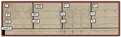
As always — I favor beginning assessment by looking first at the cardiac rhythm. For clarity — I’ve numbered the beats and labeled P waves in the long lead II rhythm strip (Figure-2):

  • The overall rhythm in Figure-2 is fairly (albeit not completely) regular — with upright P waves having a constant (and normal) PR interval in this lead II rhythm strip (RED arrows). Therefore — the underlying rhythm is sinus at a rate of ~85/minute.
  • Beat #11 is wide, of a very different morphology, and occurs early. This beat is not preceded by any P wave. Beat #11 is a PVC (Premature Ventricular Contraction).


PEARL #1: Did YOU notice that there is no P wave before beat #12? 

  • Since QRS morphology of beat #12 is identical to the QRS morphology of other sinus beats in this long lead II rhythm strip — this defines beat #12 as a junctional beat. Normal sinus rhythm then resumes with beat #13.
  • NOTE: I fully acknowledge that the fact that beat #12 is a junctional (and not a sinus) beat — does not impact at all on the management of this patient. My point is simply to emphasize that the BEST way not to overlook subtle abnormalities in the cardiac rhythm is to routinely spend the first 3-to-5 seconds of your assessment carefully surveying each beat in the long lead rhythm strip. All it takes is a 3-to-5 second look — going beat-to-beat in the long lead II rhythm strip, assessing atrial activity. In my experience — providers who fail to do this commonly miss abnormal findings in the rhythm that sometimes do make a definite difference in the management process.

Figure-2: I've numbered the beats and labeled P waves from Figure-1.


Continuing with My Systematic Approach:
After assessment of Rate and Rhythm — I favor looking next at Intervals:
  • As already noted — the PR interval is constant in front of the sinus beats, and normal in duration (I measure ~0.16 second).
  • The QRS complex is wide (ie, clearly more than half a large box in duration). Interval duration is determined by selecting that lead in which the onset and offset of the interval can be clearly seen — and in which the interval is largest. I measure QRS duration to be ~3.5 little boxes in several of the chest leads = at least 0.14 second.

PEARL #2: There are many different "systems" for the interpretation of a 12-lead ECG. That said — Regardless of which system you prefer (be it mine or any other) — it is essential to look at Intervals at an early point in the process! The reason for this — is that criteria for Axis, Chamber Enlargement and Ischemia/Infarction all change when the QRS complex is widened by BBB (Bundle Branch Block) or IVCD (IntraVentricular Conduction Defect). 
  • First establish that your patient is hemodynamically stable, and not in VT (Ventricular Tachycardia). After doing so — IF the rhythm is supraventricular, but the QRS complex is wide (as it is in Figure-2) — then STOP! Before going further — Determine WHY the QRS complex is wide!

  • In today's ECG Video Pearl (ECG-MP-22 — above) — I review my user-friendly approach for determining the reason for the conduction defect, be this RBBB (Right Bundle Branch Block) — LBBB (Left Bundle Branch Block) — or IVCD. With a little practice — You can determine if the conduction defect is RBBB, LBBB or IVCD in less than 5 seconds!

  • NOTE: In addition to today's 13-minute ECG Video — this user-friendly approach to the BBBs is reviewed in this PDF File download (excerpted from my ECG-2014-ePub).


TAKE ANOTHER LOOK at the 12-lead ECG in today's case (Figure-3):
  • We have already established that the underlying rhythm is sinus with a PVC and a junctional beat after the PVC — followed by resumption of sinus rhythm.
  • WHY is the QRS complex wide in Figure-3?

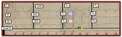
Figure-3: The 12-lead ECG and rhythm strip in today's case. What do the colored QRS complexes in leads V1, V2 and V3 tell us?


ANSWER:
The reason the QRS complex is wide in Figure-3 is that there is LBBB.
  • There is a monophasic R wave in left-sided leads I and aVL.
  • With typical LBBB — a similar all-upright R wave will usually also be seen in left-sided lead V6 — unless there is significant LVH (Left Ventricular Hypertrophy). As the left ventricle enlarges — the vector of left ventricular activity moves not only to the left, but also posteriorly. As a result — a monophasic R wave will sometimes not be seen with LBBB until more posteriorly-placed leads (such as V7, V8 or V9) are looked at. This is the case in today's tracing.
  • The other ECG criterion for LBBB (which is also satisfied in Figure-3) — is the presence of either all negative or almost all negative QRS complexes in the anterior leads.

PEARL #3: Almost all patients with LBBB have underlying heart disease. In such patients, if there is longerm hypertension, cardiomyopathy or heart failure and LBBB — the prevalence of LVH is at least 80% even before one looks at the ECG.
  • The probability that a patient with LBBB has LVH goes up to >90% — IF with LBBB there are very deep S waves (≥25-30 mm) in V1, V2 or V3. This is precisely what we see in today's tracing — in which the S waves in leads V1, V2 and V3 measure 19, 32 and 20 mm respectively (BLUE, RED and YELLOW-colored complexes in Figure-3).


QUESTION:
  • Has there been an MI? (Are Sgarbossa criteria met?).


ANSWER:
As emphasized above in today's ECG Video Pearl (ECG-MP-22), and in the accompanying PDF File download — the diagnosis of ischemia/infarction is always more challenging when there is underlying BBB.
  • For example — the ST-T wave depression seen in high-lateral leads I and aVL is to be expected with LBBB.
  • There is no primary ST elevation in today's tracing (as was seen for example, in ECG Blog #146 — and in Blog #162).


PEARL #4: In the absence of obvious acute ST-T wave changes in a patient with LBBB — use of Smith-Modified Sgarbossa Criteria may help to determine IF there is excessive discordance sufficient for diagnosis of probable acute coronary occlusion.
  • IF the J-point of the ST segment is up or down by ≥25% of QRS amplitude — then the criterion of excessive discordance is satisfied.
  • CAVEAT — It may be difficult to know where the J-point is, if anterior ST elevation with LBBB is of smooth contour (in which case you won't be able to use this criterion).
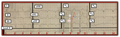
  • I've labeled today's tracing in Figure-4 — to illustrate application of Smith-Modified Sgarbossa Criteria. ST elevation is minimal in leads V1 and V3 of Figure-4. However, ST elevation is considerable in lead V2. The BLUE arrow in this figure indicates the change in slope that I take as the J-point in lead V2. The amount of J-point ST elevation is therefore 5 mm. Although lead overlap makes assessment of S wave depth difficult — the RED outline of the QRS complex in lead V2 shows the S wave to measure 32 mm. 

  • BOTTOM LINE: Although the amount of J-point ST elevation in lead V2 is considerable (ie, 5 mm) — because of marked LVH, the extremely deep S wave in this lead (of 32 mm) gives us a ratio of 5/32 — which is clearly less than 25%, such that Smith-Sgarbossa Criteria are not satisfied. 

Figure-4 Application of Smith-Modified Sgarbossa Criteria.


PEARL #5: As discussed a number of times on this blog — the ECG finding of "Fragmentation" can be insightful (See ECG Blog #165). By "fragmentation" — I mean unexpected irregularities in QRS morphology, such as notching or slurring of waveforms (as highlighted by the short, slanted YELLOW lines in no less than 6 of the leads in Figure-4).
  • Fragmentation suggests scarring — that may be the result of prior infarction, cardiomyopathy, or some other form of structural heart disease.
  • Such fragmentation does not tell us whether heart disease is acute or has been present long-term — but it nonetheless is an invaluable clue for conveying that the patient in question almost certainly has some form of underlying structural heart disease.

Finally — the Q waves in leads V4 and V5 of Figure-4 are bizarre and unexpected. This clearly is not the usual appearance of typical LBBB — in which predominant negativity in the form of either QS or rS complexes without triphasic component are usually seen in chest leads until transition to predominant positivity occurs in the lateral chest leads.


CONCLUSION: The ECG in today's case shows sinus rhythm with a PVC. There is complete LBBB with marked LVH.
  • This patient almost certainly has severe underlying heart disease — as suggested by the combination of LBBB, marked LVH + fragmentation in multiple leads + bizarre triphasic QRS complexes in leads V4 and V5, that in the context of LBBB most probably represent Q-wave equivalents. The odds are high that there have been one or more prior infarctions.
  • Although nonspecific ST-T wave abnormalities are present in multiple leads — these most probably are the result of the conduction defect, LV "strain" and underlying coronary disease. While we can not rule out the possibility of some troponin rise — there is no definitive ECG evidence on this single tracing of acute coronary occlusion. Smith-Modified Sgarbossa Criteria are negative.

 
===============================
P.S. = ADDENDUM (3/9/2024):
Some 2 years after I initially posted this case in ECG Blog #282 — I have had occasion today to review what I wrote at that time. Looking at this case today — I would amend what I said in 2022 with the following:
  • The 48-year old man presented in Blog #282 had "acute dyspnea". Additional details from the history were not provided to me. I see no indication that a repeat ECG was done. In a patient with acute symptoms (ie, shortness of breath in this case) — until such time that you can confidently either rule in or rule out an acute event — one or more repeat ECGs should be obtained. This is because development of "dynamic" ECG changes in a patient with acute symptoms could be strong evidence in support that an acute event may indeed be evolving.
  • In an acute event — evolutionary ST-T wave changes sometimes develop quickly (ie, sometimes within minutes!). As a result — that 1st repeat tracing should be obtained within ~15-20 minutes, to see if there has been ST-T wave changes during that time. This apparently was not done in this case.
  • Comparison with a prior (baseline) ECG on the patient may have been insightful. This also apparently was not done.
  •  
  • Although primary ST elevation and Smith-Modified Sgarbossa Criteria are negative in today's case — ST-T wave appearance in leads V4,V5,V6 is not what one normally expects in association with LBBB because: i) In addition to the marked QRS fragmentation in lead V4 of Figure-1 — there is uncharacteristic ST segment straightening in this lead; and, ii) There is shelf-like ST segment straightening with slight depression in leads V5,V6.
  •  
  • To EMPHASIZE: While the above cited ST-T wave findings in leads V4,V5,V6 of Figure-1 are not diagnostic — they are not "normal" findings with LBBB. As a result — additional measures would be warranted to attain more certainty as to whether or not these ST-T wave findings that I am retrospectively noting, might have been indication of an acute event (ie, Several repeat ECGs looking for "dynamic" changes should have been done — search for a prior ECG on this patient to compare with this initial ECG — and serial troponins).

  • I wish I had more follow-up on this case ... 


=======================
Acknowledgment: My appreciation to Abdullah Al Mamum (from Dhaka, Bangladesh) for the case and this tracing.
=======================


=============================
Relevant LINKS to Today's Case:
=============================

Relevant ECG Blog Posts to Today’s Post: 

  • See ECG Blog #185 — for review of the Systematic Ps, Qs, 3R Approach to Rhythm Interpretation.

  • ECG Blog #205 — Reviews my Systematic Approach to 12-lead ECG Interpretation.
  • ECG Blog #93 — Reviews at a more basic level my approach to Systematic ECG Interpretation.

  • ECG Blog #198 — An Irregular WCT (LBBB or IVCD).
  • ECG Blog #162 — LBBB with obvious STEMI.
  • ECG Blog #146 — LBBB with Acute ST-T Wave Changes.
  • ECG Blog #204 — Assessment of Sinus Rhythm and a Wide QRS (due to IVCD).

  • ECG Blog #165 — How to "Date" an Infarction — Assessment for possible LV Aneurysm — and — the diagnostic value of recognizing "Fragmentation".

  • ECG Blog #145 — RBBB with Diffuse Subendocardial Ischemia.
  • ECG Blog #144 — AV Block and Alternating BBB.
  • ECG Blog #140 — Atrial Bigeminy with Alternating RBBB/LAHB-LPHB Aberration.
  • ECG Blog #135 — Regular WCT that Looks like Fascicular VT (but is Not!).
  • ECG Blog #130 — 2nd-Degree with Alternating BBB (LBBB-RBBB).
  • ECG Blog #122 — Bifascicular Block in a Child with Congenital Heart Disease.

  • The January 31, 2022 post in Dr. Smith's ECG Blog — Reviews subtle signs of acute OMI in a patient with LBBB (Please see My Comment at the bottom of the page).
  • The June 25, 2020 post in Dr. Smith’s ECG Blog — in which I review a case of Sinus Rhythm with Intermittent RBBB.