Wednesday, December 22, 2021

ECG Blog #270 (78) — MI vs Septal Q Waves?


The ECG in Figure-1 is from a patient with chest pain, obtained several hours after cardiac catheterization.

QUESTIONS: 
  • Are there Q waves or Q wave "equivalents" in the inferior and anterior leads of ECG #1?
  • What else would YOU say about this tracing?


ANSWER: My thoughts on this ECG appear at the bottom of this post below the following "mini-didactic" on Septal vs Infarction Q waves.
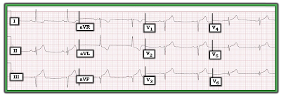
Figure-1: ECG obtained several hours after cath from a patient with chest pain. (Please note that this ECG is from the case presented in the November 20, 2019 post in Dr. Smith's ECG Blog).


================================
NOTE: I have updated this content on pointers related to Q Wave assessment from ECG Blog #94 that I wrote in 2014 — and I've added an illustrative clinical case.
  • My revised 4:30 minutes ECG Video (MP-78) on how we define "Q Waves" — and how to distinguish "normal" septal Q waves from Infarction Q waves appears further down in this post!
  • CLICK HERE — to download a PDF summary on assessment of Q Waves and for determining the ST Segment Baseline (from my ECG-2014-ePub).
  • ECG Blog #205 — Reviews my Systematic Approach to 12-Lead ECG Interpretation (including an Audio Pearl).
===============================

Assessing for Q-R-S-T Changes: Looking for Q Waves
Simply stated — a Q wave is defined as the 1st downward deflection of the QRS complex (Figure-2):
  • Small and narrow Q Waves (as seen in Panel A of Figure-2) — are often normally seen in healthy subjects. This is particularly true when such small and narrow q waves are present in one or more of the lateral leads (leads I, aVL; V4,V5,V6) — especially if the clinical scenario and the rest of the ECG appears to be benign.
  • In contrast — large and wide Q Waves (as in Panel B) — are much more likely to indicate that at some point there has been an MI (Myocardial Infarction). The larger and wider the Q waves are — and, the more leads in which they are seen — the more likely it becomes that they indicate prior MI.
  • The 3rd possibility regarding Q waves in seen in Panel C — in which there is an all-negative complex. Because there is no positive deflection (ie, no R wave)  — We are unable to tell if the all-negative deflection that is seen is: i) a Q wave; ii) an S wave; or, iii) some combination of the two. As a "compromise" — this pattern is called a QS complex.
  • NOTE: For communication and clarification purposes — we favor use of large or small-case letters for description of Q waves, depending on whether the Q wave is wide and at least 3 mm ( = 3 little boxes) deep (Figure-2).

Figure-2: Nomenclature for describing Q waves (See text).


Clinical Points Regarding Figure-2:
  • A QS pattern is most commonly seen in one or more of the anterior leads. Depending on other parameters (such as QRS complex width and ST-T wave appearance) — clinical implications of a QS pattern may be similar to clinical implications of a wide Q wave ( = possible previous infarction) — especially if QS complexes are seen in multiple leads. On the other hand — the presence of one or more QS complexes may be a normal finding (See discussion of R wave progression in ECG Blog #269).

  • PEARL #1: All bets are off! — IF QS complexes are seen in a patient with QRS widening from LBBB (Left Bundle Branch Block). In this situation — anterior QS complexes are not only common, but an expected accompaniment of LBBB (See my Review on BBB in ECG Blog #204).

  • Even when large and wide Q waves indicate that prior infarction has taken place (as would be suggested by Panel B) — the mere presence of such Q waves does not tell us anything about the "age" of the infarct (ie, whether it occurred years ago — or fairly recently).
  • To determine IF an MI might be "acute" (ie, beginning within the past few hours — and possibly still ongoing) — We use a combination of: i) History (ie, Is there new-onset chest pain? — or — Is chest discomfort more of a longterm problem without recent onset?); ii) Serial ECGs (for assessment of ST-T wave changes); iii) Cardiac markers (ie, troponin); and, iv) Other tests (ie, Echo at the bedside, looking for wall motion abnormality during chest pain — or — Cardiac Cath, as the definitive test looking for acute coronary occlusion).

  • PEARL #2: While we often think of Q waves as a longstanding marker of prior infarction — it's important to appreciate that Q waves may sometimes develop rapidly (ie, in as little as 1-2 hours after the onset of symptoms). This point emphasizes the importance of the History and comparison with prior tracings for optimal understanding of the meaning Q waves have in any particular case.

  • PEARL #3: And, although deep and wide Q waves often serve as a “marker” of prior infarction — this ECG sign is not without exceptions. Certain conditions (ie, cardiomyopathy) may produce Q waves not due to infarction (See ECG Blog #45). Further complicating assessment of Q waves on ECG — is the clinical reality that infarction-related Q waves do not always persist after the event. With time — Q waves may either decrease in size or disappear entirely. As a result — even small q waves may at times be a “marker” of prior infarction (ie, if a previously large infarction-Q-wave shrunk in size but did not disappear). Awareness of the clinical history and what the patient’s baseline ECG looked like would be needed to know if persistent small q waves in that patient were serving as a marker of prior infarction (assuming lead placement issues and a shift in axis were not the reasons for change in Q wave morphology). 
  • Bottom LINE: The term, "significant" Q wave is potentially misleading — because the size of a Q wave is not necessarily a reliable indicator of infarction. Instead of using this term — I favor describing the location and relative size of Q waves in my Descriptive Analysis — and then correlating clinically (ideally with review of prior tracings for comparison) to determine the likely significance of this finding.

==============================


ECG Media Pearl #78 (4:30 minutes Video) — ECG Blog #270 — Reviews how we define, "Q Waves" — and how to distinguish "normal" septal Q waves from Infarction Q waves.


==============================

Let's return to the case I presented at the beginning of this post.

The ECG in Figure-1 is from a patient with chest pain, obtained several hours after cardiac catheterization.

QUESTIONS that I Asked: 
  • Are there Q waves or Q wave "equivalents" in the inferior and anterior leads?
  • What else would YOU say about this tracing?


ANSWER: The ECG in today's case was taken from a patient who was found to have multi-vessel coronary disease and an acute inferior infarction. My purpose in selecting this tracing was to focus on the variety of Q-waves and Q-wave "equivalents" that are seen, which I've labeled in Figure-3. Significant ECG findings in this tracing include the following:
  • The rhythm in Figure-3 is sinus bradycardia at ~50/minute. Regarding intervals — the PR interval is upper normal (ie, ~0.20 second) — the QRS in narrow — and the QTc is normal. There is a leftward axis of about -30 degrees (isoelectric QRS in lead II — therefore not negative enough to qualify as LAHB). There is no chamber enlargement.

Regarding Q-R-S-T Changes:
  • Q waves and/or Q wave "equivalents" are seen in multiple leads.
  • Small and narrow q waves are seen in high-lateral leads I and aVL. These q waves look like normal septal q waves (because of their small size and lateral location).
  • I call the QRS complex in lead II a "Q-wave equivalent" — because for all practical purposes, clinical implications are the same as they would be for a legitimate Q wave. Note that there is an ever-so-tiny initial upward deflection to this QRS complex in lead II (RED arrow in lead II) — which is the reason this technically is not a "Q wave", but rather an rsR'S' complex. PEARL #4: In my experience, when the only thing negating the definition of a Q wave is a tiny initial positive deflection in a tracing that otherwise indicates acute infarction — this effectively serves as a marker for infarction.
  • In lead III — there is an rS complex, with a tiny-but-definitely-present initial r wave in this lead (RED arrow in lead III). By itself — this type of complex would not qualify as a "Q wave equivalent" — because unlike what we see in lead II, there is no back-and-forth change between positive and negative deflections.
  • PEARL #5: We need to remember that the ECG is the "net effect" of all ongoing electrical activity. Since the initial QRS deflections of LAHB (Left Anterior HemiBlock) and Inferior Infarction Q waves are oppositely directed — it can be extremely challenging at times to determine IF a patient has inferior lead infarction Q waves — OR — rS inferior lead complexes from LAHB — OR — some combination of the two. Therefore, in Figure-3 — although the QRS in lead II is not predominantly negative enough to qualify as LAHB, it is possible that both inferior MI and LAHB are present (ie, the QRS complexes in both leads III and aVF are very much consistent with LAHB).
  • Because of the slow heart rate — there are only 2 QRS complexes in lead aVF. The 1st QRS complex in this lead shows a definite small-but-present initial r wave, followed by a notched (fragmented) descent of the S wave (RED arrow in lead aVF). However, I do not see any initial positive deflection in the 2nd QRS complex in lead II — which qualifies this complex as a QS in lead aVF.
  • PEARL #6: Regardless of whether true QRS morphology in lead aVF is an rS or QS complex — I call the "slurring" in the initial S wave descent in this lead (RED arrow in lead aVF) as a "Q-wave equivalent" — since in the context of other ECG findings of acute infarction, this "slurring" in an inferior lead serves as a marker for infarction.
  • PEARL #7: Small and narrow q waves are seen in leads V5 and V6 (GREEN arrows in these leads). That said — by the concept of proportionality — these q waves are disproportionately large considering the tiny size of the R wave in leads V5 and V6. Especially in view of associated ST-T wave changes — despite their tiny size — I'd call these "infarction q waves".

Figure-3: I've labeled the initial tracing in today's case (See text).


Regarding R-Wave Progression:
  • There is poor R-Wave Progression (PRWP) in the chest leads of Figure-3 — because Transition is delayed, and does not occur until leads V5-to-V6, if then (ie, the R wave in lead V6 is barely more positive than the S wave in this lead is deep — See ECG Blog #269 for review of R Wave Progression).
  • Note that although it looks as if there is a QS complex in lead V3 (BLUE arrow) — there is actually a small, initial positive deflection (r wave) to the QRS complex in this lead. Since leads V1, V2 and V3 are simultaneously recorded — the vertical dotted BLUE line in Figure-3 defines the onset of the QRS in these 3 leads.
  • PEARL #8: Although lead V3 does not manifest a QS complex — there is "Loss of R-Wave" from lead V2-to-V3 — which in the context of the acute ST-T wave changes, serves as a "Q-wave equivalent" marker of anterior infarction!

Regarding ST-T Wave Changes:
  • Hyperacute T-waves (that are taller, fatter-at-their-peak, and wider-at-their-base than expected) are seen in multiple leads (ie, in the 3 inferior leads — and in all 6 chest leads). Virtually all of these leads show at least slight J-point ST elevation (which attains ~1.5 mm of ST elevation in each of the inferior leads — See ECG Blog #218 for more on Hyperacute T waves).
  • There is ST-T wave depression in high-lateral leads I and aVL in Figure-3. Note the mirror-image opposite relationship for the ST-T wave depression in lead aVL (including 1-1.5 mm of J-point depression) — compared to the shape of the ST elevation and hyperacute T wave in lead III. In the setting of new chest pain — this ECG finding is diagnostic of acute inferior infarction (See ECG Blog #184 for more on this topic).

Putting It All Together:
My goal in writing today's ECG Blog #270 — was to focus on the evaluation of Q waves and Q wave "equivalents" by means of didactic review and an illustrative case. Given the history of chest pain — the ECG in Figure-3 is diagnostic of acute infarction. ECG findings of concern include:
  • Fairly marked sinus bradycardia.
  • Signs of acute inferior infarction — including Q waves or Q wave "equivalents" in leads II and aVF — inferior lead ST elevation and hyperacute T waves — mirror-image opposite ST-T wave depression in lead aVL.
  • Suggestion of acute antero-lateral infarction — including "loss" of R wave (from lead V2-to-V3) — disproportionately large q waves in leads V5 and V6 — hyperacute T waves in all 6 chest leads.


===============================
Related Material of Interest: 
  • CLICK HERE — to download a PDF summary on assessment of Q Waves and the ST Segment Baseline (from my ECG-2014-ePub).

  • ECG Blog #205 — Reviews my Systematic Approach to 12-Lead ECG Interpretation (including an Audio Pearl).

  • ECG Blog #269 — Reviews assessment of R Wave Progression (including the causes of "Poor" R wave progression).
  • ECG Blog #204 — Reviews the ECG diagnosis of Bundle Branch Block (RBBB, LBBB, IVCD).
  • ECG Blog #45 — Reviews the unusual ECG findings in this patient with Muscular Dystrophy (which is known for the occurrence of cardiomyopathies).
  • ECG Blog #218 — Reviews what is a Hyperacute T Wave (including the Audio Pearl in this post ).
  • ECG Blog #184 — Reviews the "magical" mirror-image opposite relationship with acute infarction between lead III and lead aVL (including the Audio Pearl in this post).

  • Please note that the ECG shown in Figure-1 of today's case is from the November 20, 2019 post in Dr. Smith's ECG Blog. (This is ECG #5 from my figure in My Comment at the BOTTOM of the page in that Dr. Smith post).


Saturday, December 18, 2021

ECG Blog #269 (77) — What is Poor R Wave Progression? Why Care?


================================
NOTE: I have updated this content on assessment of R Wave Progression from ECG Blog #94 that I wrote in 2014.
  • My revised 11-minute ECG Video (MP-77) on assessment of R Wave Progression appears at the BOTTOM of this post!
  • CLICK HERE — to download a PDF summary on assessment of R Wave Progression (from ECG-2014-ePub).
  • ECG Blog #248 — Reviews the Causes of a Tall R in Lead V1 (including the Audio Pearl in this blog post).
  • ECG Blog #205 — Reviews my Systematic Approach to 12-Lead ECG Interpretation (including an Audio Pearl).
===============================

Assessing “R Wave Progression"
Our purpose in using “Q-R-S-T” as a memory aid — is so as not to forget to evaluate the “R” component = “R” Wave Progression in the precordial leads. We schematically illustrate this concept in Figure-1 — in which the heart’s electrical activity is depicted in the Transverse (Horizontal) Plane. The predominant direction of this electrical activity is to the left and posteriorly (as suggested by the large RED arrow in Figure-1). This predominant direction of electrical activity accounts for the change in appearance of the QRS complex in precordial leads V1-thru-V6 as we move across the chest:
  • Initially in right-sided lead V1 — the QRS is predominantly negative (an rS complex). This is because lead V1 sees most of the heart’s electrical activity as moving away from the right as the much larger and thicker LV (Left Ventricle) is depolarized.
  • Note how the R Wave “progresses” — by getting taller as one moves from leads V1 and V2 toward left-sided leads V5 and V6. In general — R wave amplitude “tops out” by lead V5, and then becomes smaller in lead V6. This is the situation in Figure-1 — although many variations on this pattern of R wave progression are possible.
  • We define the point of “Transition” — as the place where the R wave becomes taller than the S wave is deep (highlighted by the GREEN arrow in Figure-1). Normally — transition occurs between leads V2-to-V4. It occurs between lead V3-to-V4 in Figure-1.

Figure-1: Schematic blow-up cross-section of the heart. Precordial lead placement of recording electrodes is suggested by the YELLOW ovals. Note our focus on the predominant direction of the heart’s electrical activity during ventricular depolarization (RED arrow) — and the effect this has on progression of R wave amplitude in the precordial (chest) leads. Initially (ie, in leads V1,V2) — there is a predominant S wave (negative deflection) — because most of the heart’s electrical activity is moving away from these right-sided leads. As one moves across the chest — the R wave becomes taller until the point of transition is reached where the R wave becomes taller than the S wave is deep (GREEN arrow). This occurs between lead V3-to-V4 in this Figure (Transition normally occurs somewhere between lead V2-to-V4).


Septal Depolarization: The final point illustrated in Figure-1 — is the effect septal depolarization may have on the ECG. After the sinus-conducted impulse leaves the AV Node — the very first part of the ventricles to be depolarized is the left side of the interventricular septum. As a result — septal activation normally proceeds from left-to-right (small BLUE arrow in Figure-1).
  • Because septal activation normally moves toward right-sided lead V1 — there will often be a small initial r wave (positive deflection) in lead V1, as well as in lead V2 (as is seen in these leads in Figure-1). This small initial r wave may be lost when there is septal infarction.
  • At the same time — septal activation normally moves away from left-sided leads V5,V6. As a result — one or more lateral leads (ie, leads I,aVL; V4,V5,V6) will often manifest a small initial q wave (negative deflection) simply as a reflection of normal septal activation. 
  • PEARL #1: Lateral q waves are unlikely to indicate prior infarction IF: i) These q waves are small and narrow; and, ii) The clinical scenario and the rest of the ECG appear to be benign.

========================

TEST YOURSELF: Where is the Point of Transition?
Look at the 3 schematic precordial lead sequences in Panels A, B and C of Figure-2. Comment on R wave progression in each of these sequences.
  • In each case — Indicate the 2 leads between which Transition occurs.
  • What do you think is meant by the term, “late” Transition?

Figure-2: Identify the point of Transition for the 3 precordial lead sequences shown in this Figure. What is meant by the term, "late" Transition? HINT: Transition normally occurs somewhere between lead V2-to-V4.


ANSWER to Figure-2: The R wave “progresses” in each of the 3 sequences shown in this Figure. By this we mean that R wave amplitude is smallest in lead V1 — and then gets taller as one moves to the next precordial leads.
  • Normal Transition occurs in Panel B. That is — the R wave becomes taller than the S wave is deep between lead V3-to-V4.
  • In Panel A — there is “early” Transition. By this we mean that the R wave becomes taller than the S wave is deep before the normal point of transition. Specifically — Transition in Panel A occurs between V1-to-V2.
  • In Panel C — there is "late" Transition. That is, the R wave becomes taller than the S wave is deep after the normal point of transition. Specifically — Transition in Panel C occurs between V4-to-V5.

Putting It Together: Assessing “R Wave Progression"
Insight into the clinical utility of routinely assessing the "R" component in the "Q-R-S-T" parameter is forthcoming from review of Figure-3.
  • Panel A in Figure-3 — shows normal R wave progression. Note progressive increase in R wave amplitude across successive precordial leads until the R wave “tops out” in lead V5. Transition is normal in Panel A (occurs between lead V3-to-V4). 
  • PEARL #2: Although a QS complex is present in lead V1 — this is unlikely to reflect prior septal infarction because the QS complex is limited to lead V1 (ie, an r wave develops by lead V2 in Panel A). The small and narrow q waves in leads V5,V6 almost certainly represent normal septal q waves. They are unlikely to indicate prior infarction — because of their small size and expected lateral location.

  • In contrast — anterior infarction is suggested by the precordial lead sequence in Panel B. This is because a QS complex is seen not only in leads V1,V2 — but also in lead V3. FINE Points: We can more specifically qualify location of the infarct in Panel B as being “antero-septal” — because there is loss of the initial r wave in leads V1,V2 (ie, loss of the initial r wave in lead V1 suggests "septal" involvement). 
  • Note in Panel B, that despite the QS in V1-thru-V3 — transition is still “normal” (occurs between lead V3-to-V4). Finally — the isolated small and narrow q wave in lead V6 of Panel B is unrelated to the anterior infarction — and almost certainly once again reflects a normal septal q wave.

  • In contrast, in Panel C — a QS pattern is seen in both lead V1 and V2, but not in lead V3. That is — a small-but-definite initial positive deflection (r wave) is seen in lead V3. The finding of a QS complex in V1,V2 but not in V3 — may or may not be a "normal" finding. Instead, it could indicate: i) A technical lead placement error: ii) Septal infarction; or, iii) A normal variant. Since there may not be any reliable way to distinguish between normal variant vs lead placement error vs prior septal infarction — We simply acknowledge, "QS in V1,V2 — Suggest clinical correlation" in our interpretation. 
  • PEARL #3: Statistical odds are far greater (approximately 4-to-1) that either normal variant or lead placement error rather than septal infarction is the reason for a QS complex in V1,V2 but not V3 (as seen in Panel C).

Figure-3: Normal RWP (R Wave Progression) is seen in Panel A. This is in comparison with precordial lead sequences suggestive of anterior MI (Panel B) vs an indeterminate pattern showing a QS complex in leads V1,V2 but not in V3 (Panel C).


KEY Point: The above comments regarding clinical implications of a QS complex in leads V1,V2,V3 hold true only when the QRS complex is narrow. 
  • As we have already noted — QRS widening due to LBBB commonly produces QS complexes in anterior leads that are simply the result of the conduction defect (and are not indicative of anterior infarction!).

PEARL #4: There are at least 3 other common reasons for PRWP (Poor R Wave Progression) with a narrow QRS complex that are not the result of prior infarction. These are: i) LVH; ii) LAHB; and, iii) Severe pulmonary disease/COPD.
  • Usually at least some r wave will be seen by lead V3 with these other conditions — though not always. 
  • Bottom Line: It is sometimes extremely difficult to know with certainty the cause of PRWP when R waves in leads V1,V2,V3 are either very small or absent. Clinical correlation is essential. (See Figure-4 below for a full list of causes of PRWP).

B
eyond-the-Core: It is insightful to appreciate the physiologic reason why R wave progression is often “poor” in the presence of these other conditions.
  • LVH (Left Ventricular Hypertrophy) — produces an increase in leftward and posterior forces. When marked — these leftward and posterior forces may totally predominate over initial rightward and anterior forces seen in leads V1,V2,V3. The result is reduction in r wave amplitude in anterior leads. At times — there may even be elimination of any r wave at all in leads V1,V2,V3.
  • LAHB (Left Anterior HemiBlock) — The bundle of conduction fibers in the LAH (Left Anterior Hemifascicle) lies slightly in front of (anterior to) fibers in the LPH (Left Posterior Hemifascicle). As a result — block in the LAH (as occurs with LAHB) results in an initial posterior direction for electrical activity that is first conducted over the intact LPH. The result may once again be reduction in r wave amplitude in the anterior leads.
  • COPD (Chronic Obstructive Pulmonary Disease) — Severe longstanding pulmonary disease leads to an increase in right ventricular forces. In addition — orientation of the heart’s electrical forces in 3 dimensions may be altered by the large emphysematous chest of a patient with chronic COPD. These physiologic changes may result in ECG signs of: i) Poor R wave progression; ii) Persistence of relatively deep precordial S waves in lateral chest leads; and, iii) Reduced R wave amplitude across the chest leads. In the extreme — R waves are markedly reduced in size (and may even be absent) in anterior precordial leads.

==================================


ECG Media Pearl #77 (11:00 minutes Video) — Reviews assessment of R Wave Progression as part of the Q-R-S-T memory aid in the Systematic Approach to 12-lead interpretation (includes clarification of the term, "Poor R Wave Progression vs Transition).


Figure-4: Causes of PRWP (See the Video Pearl just above).

===============================

Related Material of Interest: 
  • CLICK HERE — to download a PDF summary on assessment of R Wave Progression (from the ECG-2014-ePub).
  • ECG Blog #248 — Reviews the Causes of a Tall R in Lead V1 (including the Audio Pearl in this blog post).
  • ECG Blog #205 — Reviews my Systematic Approach to 12-Lead ECG Interpretation (including an Audio Pearl).


Sunday, December 12, 2021

ECG Blog #268 (76) — Mobitz I vs Complete AV Block vs Supernormal Conduction?


The ECG in Figure-1 was obtained from a 54-year old man, who presented to the ED (Emergency Department) with chest pain. There is obvious inferior MI and AV block — but from a teaching perspective — there is so much more!

  • In addition to inferior MI — What other areas of the heart are affected?
  • What is the probable age of the infarction?
  • What is the rhythm? Is this Mobitz I? — Complete AV block? — or something else? 


Figure-1: 12-lead ECG and 2-lead rhythm strip obtained from a 54-year old man with chest pain.


=============================

NOTE #1: I am indebted to Dawn Altman, RN, EMT-P — whose ECG Guru web site has been posting ECGs and Arrhythmias available for teaching purposes since 2011. My appreciation to Dawn for allowing me to expand on this case — that she originally posted on the ECG Guru.

=======================================

NOTE #2: Some readers may prefer at this point to listen to the 3:10 minute ECG Audio PEARL before reading My Thoughts regarding interpretation of the rhythm in today's case. Feel free at any time to review to My Thoughts on this tracing (that appear below ECG MP-76).

=======================================


Today’s ECG Media PEARL #76 (3:10 minutes Audio) — Briefly reviews the concept of "Supernormal" Conduction — in which conduction for a brief period in the cardiac cycle is better-than-thought-to-be possible.


MY Sequential Approach to the ECG in Figure-1:

There is a series of important ECG findings in this case — and the cardiac rhythm is fascinating. Although I used a Systematic Approach (as I always do) to interpret both the cardiac rhythm and the 12-lead ECG (See ECG Blog #185 and Blog #205 for details on my approach) — I did allow myself the "luxury" of a few seconds to take in the principal findings. Beginning with the 12-lead ECG — I'd make the following points:

  • Clearly there has been recent inferior MI, with obvious ST elevation in each of the inferior leads. But what is the AGE of this inferior infarction? There are small-but-present q waves in leads II and aVF — with already a large Q wave in lead III. 
  • Not only is there marked ST elevation in lead III — but there is beginning T wave inversion, signaling the likely onset of reperfusion T waves.
  • The ST-T wave in lead aVL is almost the mirror-image reciprocal picture of the ST-T wave in lead III. This confirms that the inferior MI is at least "recent" (See Figure-2). That said — the unexpectedly tall terminal T wave in lead aVL (which in its "mirror-image" corresponds to T wave inversion) confirms the presence of what we saw in lead III = reperfusion T waves. Spontaneous reperfusion (without thrombolytics or angioplasty) is common during the course of acute MI — and this is undoubtedly ongoing in today's case.
  • There is also associated acute posterior infarction — as signaled by the positive "Mirror Test" picture for the ST-T waves in anterior leads, which is maximal in leads V2 and V3 (See Figure-2). Further support of associated posterior MI is forthcoming from the taller-than-expected R waves in leads V2 and V3 (which in its "mirror-image" corresponds to increasingly deep Q waves).
  • There is almost certainly also acute RV (Right Ventricular) infarction — as suggested by minimal ST depression in lead V1 in the face of maximal downsloping ST depression in leads V2,V3 (See ECG Blog #190). This could be confirmed by obtaining right-sided leads. That said — Lead V1 is in effect, a "right-sided" lead — and in the setting of acute proximal RCA (Right Coronary Artery) occlusion — there will often be infero-postero and RV infarction, as a result of the anatomic distribution of the RCA. 
  • On ECG — the effect of simultaneous ST elevation in lead V1 (from acute RV MI) — together with the ST depression in lead V1 from acute posterior MI — will often yield a "net effect" of a nearly flat ST segment in lead V1. In neighboring leads V2, V3 (which are not "right"-sided) — we don't see any effect from the ongoing acute RV MI, and as a result there is much greater ST depression in other anterior leads. In contrast, with acute infero-postero MI, but in the absence of acute RV MI — significant ST depression will usually also be seen in lead V1, as well as other anterior leads. 
  • NOTE: Although in Figure-1 there is T wave inversion in lead V1 from reperfusion — the amount of ST depression in this lead is modest compared to the amount of ST depression in leads V2 and V3.
  • NOTE: As expected with posterior infarction, there is less ST depression in lateral chest leads (ie, leads V5 and V6). That said — tall T waves with marked T wave peaking persist through to lead V6. Although I would definitely check serum K+ — I suspect this picture is not due to hyperkalemia — but rather to extension of marked "mirror-image opposite" reperfusion T waves. Note that the History we were given did not specificy the time symptoms began. Given the large Q wave in lead III — the taller-than-expected R waves in leads V2,V3 — and especially given diffuse and marked suggestion of reperfusion T waves — I'd suspect that although this extensive MI is "recent" — it is less likely to have just begun in the past couple hours (More history would be needed to better determine the likely time of onset of the infarct).
  • Final NOTE: Although chest lead T waves are tall and peaked — this probably is not a manifestion of deWinter T waves from acute LAD (Left Anterior Descending) coronary artery occlusion because: i) There are very prominent changes of acute inferior MI in the limb leads; ii) I'd expect a different ST-T wave appearance in lead V1 if the infarction was the result of acute LAD occlusion; and, iii) The duration of depressed ST segments in the chest leads is relatively long (whereas with deWinter T waves, the slightly depressed J-point ST depression much more rapidly rises to form giant T waves than what we see in today's tracing — as is discussed in more detail in ECG Blog #266).


Figure-2: I focus on the 12-lead ECG in today's case — to highlight the "magical" mirror-image-opposite relationship between lead III and lead aVL (which confirms recent inferior infarction). Application of the "Mirror Test" to the QRST complex in leads V2 and V3 confirms associated posterior MI (See text).


What about the Rhythm in Today's Case?

I do not think we have a sufficient period of ECG monitoring for definitive interpretation of today's rhythm. That said — I believe we can define the principal rhythm abnormality, and speculate on the fascinating possibilities of a few unique findings.

  • For clarity — I reproduce the simultaneously-recorded 2-lead rhythm strip in Figure-3. By the Ps, Qs & 3R Approach — the QRS is narrow — a certain number of P waves are definitely present — and, the rhythm is almost, but not completely Regular (with a Rate of ~55/minute after beat #3). 

Figure-3: I focus on the rhythm strip in today's case (See text).


This initial rhythm assessment leaves us with 3 QUESTIONS:

  • Are P waves regular throughout the rhythm in Figure-3?
  • IF P waves are regular — Are we seeing a 3:2 Mobitz I cycle ( = AV Wenckebach) at the beginning of the tracing?
  • After beat #3 — Are P waves Related to neighboring QRS complexes?


Are P Waves Regular throughout the Rhythm?

The challenge in assessing P wave regularity in today's case — is that many of the P waves are partially hidden within the ST segment.

  • Calipers provide invaluable assistance.
  • Find 2 P waves in a row that you are certain about. I chose the first 2 RED arrow P waves in Figure-4.
  • Setting your calipers to the P-P interval of these first 2 RED arrow P waves — you can now "walk out" regular P wave deflections throughout the entire rhythm strip (RED arrows in Figure-4).


Figure-4: Regular P waves are seen throughout this tracing (RED arrows).


My Initial Laddergram:

For those interested in a "primer" on how to read (and/or draw) Laddergrams — Please check out ECG Blog #188.

  • My initial proposed laddergram for the mechanism of the rhythm in today's case is shown in Figure-5. This suggests a 2 beat grouping at the beginning of the tracing of Mobitz I, 2nd-degree AV block with 3:2 Wenckebach conduction for beats #1 and #2 (PURPLE lines within the AV nodal tier in Figure-5).
  • Continuing with the laddergram — There appears to be complete (3rd-degree) AV block, beginning with beat #3. ARROWS in the lead II rhythm strip show the occurrence of a regular atrial rhythm at the surprisingly fast rate of 155/minute. Whether this represents marked sinus tachycardia vs an ectopic ATach (Atrial Tachycardia) is unknown from this single single tracing.
  • None of the P waves after beat #3 in Figure-5 conduct. The way we know this — is that although the narrow-QRS ventricular rhythm is perfectly regular after beat #3 (at a rate of ~55/minute) — the PR interval continually changes (If you focus on each PINK arrow P wave before the QRS of beats #4, 5, 6, 7, 8 and 9 — Caliper measurement confirms a small-but-definitely-present continual change in the PR interval!). This means that there is complete AV dissociation — because after beat #3, none of the P waves conduct! I suspect this represents complete AV block — although additional monitoring would be needed to confirm this.


As an Advanced Point — it is difficult to guarantee that all P waves fail to conduct in Figure-5 despite being given adequate opportunity to do so (as would be required if there truly was complete AV block). This is because: i) The ventricular rate is faster than 50/minute; and, ii) Because the atrial rate is so fast. 

  • PEARL #1: Complete AV block is most reliably diagnosed when the ventricular rate is slow enough to guarantee that all P waves have adequate opportunity to conduct — yet still fail to do so (which optimally requires a ventricular rate of ~50/minute or less).
  • PEARL #2: Given rapidity of the atrial rate at ~155/minute (and the difficulty of distinguishing between a very rapid sinus tachycardia vs ectopic ATach) — it's impossible to know how much AV conduction might improve if the atrial rate were to slow down.


Despite these reservations (and assuming the rhythm after beat #3 truly is complete AV block) — this raises the question of HOW can you have Mobitz I just 1 beat before you develop complete AV block?

  • While hard to say that the mechanism I initially proposed in Figure-5 is "impossible" — such an abrupt transition from 2nd-degree Mobitz I — to complete AV block in the space of a single beat — seems highly improbable! As a result, on further reflection — I did not think this initial laddergram represented a plausible mechanism for today's rhythm.


Figure-5: My initial proposed laddergram for the mechanism of today's rhythm (See text).


My Revised Laddergram:

On further reflection — I revised my initial laddergram to the laddergram shown in Panel B of Figure-6. For the purpose of comparison — I show both laddergrams together in this Figure.

  • The reason I do not think beats #1 and #2 represent a 3:2 Mobitz I grouping — is that the PINK arrow P wave just before beat #9 appears to have the same PR interval as for the P wave before beat #1 — yet we have already established that the P wave before beat #9 is not conducting (because of the presence of complete AV dissociation).
  • The "good news" about this 2nd laddergram that I propose in Panel B — is that instead of having to explain the path of several P waves as they pass through the AV Nodal Tier (as per the several PURPLE lines in Panel A) — I only have to explain the path of 1 P wave ( = the RED arrow in the ST segment of beat #1). I suspect that the reason this 1 P wave in Panel B is able to conduct to the ventricles — is a result of "supernormal" conduction! This controversial phenomenon is defined as "better-than-expected conduction" for a brief period during the cardiac cycle (See today's Audio Pearl — and the article by Elizari et al).
  • I fully acknowledge the controversy surrounding the concept of "supernormal" conduction in humans. Despite me being aware of this described phenomenon for several decades — I count on the fingers of one hand the number of times that I thought I truly detected it. But for today's case, in which there is extensive recent inferior MI, with what appears to be complete AV block with a junctional escape rhythm — I find theorizing "supernormal" conduction to be the most LOGICAL way to explain the early occurrence of beat #2. The P wave in the ST segment of beat #1 is located at a spot in the cardiac cycle where it would seem most likely to encounter that "magic point" when depressed conduction might be transiently facilitated.
  • KEY Point: What I especially like about postulating the occurrence of "supernormal" conduction as the mechanism for explaining why early beat #2 is conducted — is that this supports my contention that it is all-but-impossible for a short grouping of Mobitz I to abruptly interrupt complete AV block in the space of a single beat.


Figure-6: My revised proposed laddergram for the mechanism of today's rhythm is shown in Panel B. For comparison — I've reproduced my initial laddergrm in Panel A. Given how unlikely it would seem to abruptly change from Mobitz I to complete AV block in the space of a single beat — I feel my laddergram in Panel B is a much more logical proposal (See text).


==================================

Acknowledgment: My appreciation to Dawn Altman of the ECG Guru (Dawn lives in Tennessee, USA) for allowing me to use this tracing and this case.

==================================


Relevant ECG Posts to Today's Case: