Saturday, January 8, 2022

ECG Blog #274 — New or Old Septal Infarction?


The ECG in Figure-1 was obtained from an older woman with known coronary disease — who presented with shortness of breath and some chest discomfort.


QUESTIONS:

  • How would YOU interpret the ECG in Figure-1?
  • Is there evidence of new or old septal infarction?
  • WHY do you think I am presenting this case?


Figure-1: ECG from an older woman with known coronary disease — with shortness of breath and some chest discomfort.


=======================================

NOTE: Some readers may prefer at this point to watch to the 11:00-minute ECG Video PEARL before reading My Thoughts regarding the ECG in Figure-1. Feel free at any time to refer to My Thoughts on these tracings (that appear below ECG MP-77).

=======================================


ECG Media Pearl #77 (11:00 minutes Video) — Reviews assessment of R-Wave Progression as part of the Q-R-S-T memory aid in the Systematic Approach to 12-lead interpretation (includes clarification of PRWP vs delayed Transition).

  • Common causes of PRWP (Poor R Wave Progression) are shown below in Figure-2. The fact that conditions as opposite as LVH and RVH may cause "PRWP" — calls into question the clinical utility of this designation. For this reason — I favor simply describing what is seen regarding chest lead QRS appearance, including the zone of Transition (the leads between which the R wave becomes taller than the S wave is deep).
  • For more on assessment of R-Wave Progression — Please see ECG Blog #269.


Figure-2: Common causes of PRWP (See the Video Pearl in today's case!).


My THOUGHTS on ECG #1:

As always — I approached the ECG shown in Figure-1 systematically (See ECG Blog #205 for my Systematic Approach). My interpretation was as follows: 

  • Sinus rhythm @80/minute.
  • Normal PR and QTc intervals. The QRS appeared to be slightly wide (ie, ~0.11 second) — though not in a bundle branch block pattern.
  • There is marked left axis deviation — consistent with LAHB (Left Anterior HemiBlock). As reviewed in ECG Blog #203 — diagnosis of LAHB can usually be made within seconds by appreciation of a predominantly negative QRS complex in lead II, in association with a positive QRS in lead I. NOTE: LAHB may prolong the QRS by 0.01-0.02 second — so this could account for the slight QRS widening.
  • No chamber enlargement.


Regarding Q-R-S-T Changes:

  • Q waves: Small and narrow q waves are seen in lateral leads I and aVL. Their appearance is consistent with normal septal Q waves. Potentially not so benign are the Q waves seen in leads V1 and V2.
  • R wave progression: Transition (ie, the point where the R wave gets taller than the S wave is deep) is either delayed or never occurs in ECG #1 (ie, it's difficult to determine if the R wave in lead V6 is or is not greater than the S wave in this lead). This qualifies as PRWP (Poor R Wave Progression).
  • ST-T wave changes: There is nonspecific ST-T wave flattening in multiple leads, that overall does not look acute. That said — What about the positive T wave in lead V2 that potentially is disproportionately tall considering how tiny the QRS complex is in this lead? 


So WHY is there PRWP?

As discussed in today's Video Pearl (and as shown above in Figure-2) — there are a variety of potential causes of PRWP. The most relevant potential causes to consider in today's case include:

  • LAHB.
  • Pulmonary disease.
  • Lead misplacement.
  • Anterior or anteroseptal infarction.


Why might LAHB (Left Anterior HemiBlock) cause PRWP? 

  • The bundle of conduction fibers in the LAH (Left Anterior Hemifascicle) lies slightly in front of (anterior to) fibers in the LPH (Left Posterior Hemifascicle). As a result — block in the LAH (as occurs with LAHB) results in an initial posterior direction for electrical activity, that is first conducted over the intact LPH. This often leads to reduction in r wave amplitude in the anterior leads — which may sometimes simulate anterior infarction.


What about COPD (Chronic Obstructive Pulmonary Disease)?

  • Severe longstanding pulmonary disease leads to an increase in right ventricular forces. In addition — orientation of the heart’s electrical forces in 3 dimensions may be altered by the large emphysematous chest of a patient with chronic COPD. These physiologic changes may result in ECG signs of: i) Poor R wave progression; ii) Persistence of relatively deep precordial S waves in lateral chest leads; and, iii) Reduced R wave amplitude across the chest leads. In the extreme — R waves are markedly reduced in size (and may even be absent) in anterior precordial leads (See ECG Blog #234 for review on the ECG diagnosis of pulmonary disease and RVH).


WHY am I Presenting this CASE?

The principal reason I chose to present this case — is to highlight lead misplacement as the most likely cause for the Q waves in leads V1 and V2 of Figure-1.

  • Chest lead misplacement of leads V1 and V2 is surprisingly common. In an informal study I participated in, in which a series of internet-published examples supposedly illustrating "proper lead placement" were reviewed — more than half of these published examples showed faulty lead placement. That is — instead of proper placement of the lead V1 and V2 electrodes, just to the right and to the left in the 4th ICS (InterCostal Space), as shown in Figure-3 — electrode placement of these 2 leads was in the 3rd, or even as highly placed as the 2nd ICS! Given this frequency of errors among published "teaching examples" — One can only wonder how widespread faulty placement of leads V1 and V3 must really be in clinical practice!
  • In my experience over the past decade in reviewing and commenting on acute ECGs posted on numerous international ECG internet forums — emergency care providers commonly fail to recognize this form of lead misplacement. 


Figure-3: Anatomic landmarks for placement of the 6 precordial leads (from Grauer K: ECG-2014-ePub). Note that leads V1 and V2 should be placed in the 4th ICS (InterCostal Space), just to the right and to the left of the sternum. All too often — these leads are placed too high on the chest (in the 2nd or 3rd ICS).


===========================

3 Clues to Quickly Recognize V1, V2 Misplacement:

Despite the alarming frequency of lead placement errors — the "good news" is that it is EASY to suspect when misplacement of leads V1/V2 is likely! There are 3 clues to look for:


Suspect leads V1 and/or V2 have been placed 1 or 2 interspaces too high on the chest IF one or more of the following clues are present:

  • Clue #1 — If there is an r’ in leads V1 and/or V2, especially if the other finding for incomplete RBBB (ie, terminal s waves in lateral leads I and V6) is absent.
  • Clue #2 — If there is a significant negative component to the P wave in lead V1 and/or V2.
  • Clue #3 — If the appearance of the P wave, QRS complex and ST-T wave in leads V1 and V2 looks very much like the P wave, QRS complex and ST-T wave in lead aVR.


NOTE: As helpful as the above 3 Clues are for quickly suspecting too high placement of leads V1 and/or V2 — these clues are not infallible. There are times when despite our suspicion from looking at an ECG that leads V1 and V2 have been placed too high — this will turn out not to be the case. For example: i) Incomplete RBBB may truly be present (resulting in a terminal r' in leads V1 and/or V2); ii) An all negative, relatively large P wave may sometimes be seen in leads V1,V2 as a normal finding — or — the patient may have true left atrial enlargement; iii) Variation in body habitus and chest wall anatomy may produce unusual ECG findings; and/or, iv) The patient may truly have had prior septal infarction.

  • Therefore, my goal in presenting these 3 clues — is that they suggest when to suspect that an unusual anterior lead appearance might best by explained by too-high positioning of lead V1 and/or V2.
  • It's EASY to check out your suspicion. Simply verify that chest lead placement is correct — and then REPEAT the ECG!


===========================


TRY OUT these 3 Clues:

ECG "B" in Figure-4 was obtained just a short while after ECG "A".

  • WHY did the treating clinician suspect too-high placement of leads V1 and V2 in ECG "A"?
  • What changes do you see in the repeat ECG ( = ECG "B") after correcting for erroneous placement of leads V1 and V2?


Figure-4: Comparison of "before" and "after" tracings — to illustrate ECG findings of too-high placement of the lead V1 and V2 electrodes. (NOTE: I've adapted the 2 ECGs in this Figure from the November 4, 2018 post in Dr. Smith's ECG Blog). 


ANSWERS:

The clinical concern on seeing ECG "A" in Figure-4 — was whether the Q waves in leads V1 and V2 indicated prior septal infarction? The reason too-high placement of the V1 and V2 electrodes was suspected — is that all 3 Clues described above are seen:

  • There is a significant negative component to the P waves in leads V1 and V2 (these P waves are all negative in ECG "A").
  • There is a small-but-definite terminal r' after the large Q waves in both leads V1 and V2.
  • The P wave, QRS and T wave appearance in lead V1 closely resembles the appearance of these deflections in lead aVR.
  • In addition — there is a rather abrupt transition from the tiny QRS complexes in leads V1,V2 — to the significantly larger QRST complex with positive P wave in lead V3.


After correcting for erroneous V1,V2 lead placement — ECG "B" now shows:

  • Restoration of positive P waves in both V1 and V2 (with the positive P wave in lead V2 now closely resembling P wave morphology in neighboring lead V3).
  • Disappearance of the small-but-present terminal r' in leads V1 and V2.
  • A completely different P-QRS-T wave appearance for leads V1 and V2 — compared to lead aVR (See Figure-5 for a magnified view of the changes in leads V1,V2 after correction of erroneous lead placement).
  • NOTE: I suspect that the position of lead V3 in ECG "A" of Figure-4 may have also been incorrect — since after checking lead placement position and repeating the ECG — there is now (in ECG "B") a predominantly positive QRS complex in lead V3.


Figure-5: Magnified view of leads aVR, V1 and V2 from Figure-4 — to emphasize ECG findings before and after correcting for too-high placement of leads V1 and V2. Note disappearance of the terminal r' (BLUE arrows in ECG "A") — positivity of the P waves in leads V1,V2 in ECG "B" — and loss of resemblance between the P-QRS-T wave appearance of leads V1,V2 with lead aVR.


Today's CASE Conclusion:

Let's return to the initial ECG in today's case (Figure-6). I previously listed the 4 conditions most likely to account for the PRWP in ECG #1. These were:

  • LAHB.
  • Pulmonary disease.
  • Lead misplacement.
  • Anterior or anteroseptal infarction.


Armed with the 3 Clues for suspecting leads V1,V2 misplacement — Let's now revisit these 4 entities:

  • LAHB is present in ECG #1 (See Figure-6). Therefore — this conduction defect may indeed be contributing to the PRWP and lack of initial r wave in leads V1 and V2. The slight QRS widening and presence of LAHB in this tracing could both be the result of this patient's known coronary disease.
  • Although we are only provided with limited information about the medical history of today's patient — we are told that this older woman presented with shortness of breath. Poor R wave progression — marked frontal plane axis deviation — and persistence of sizeable S waves through to lead V6 can all be seen with chronic pulmonary disease. That said — other ECG findings of pulmonary disease are not seen in ECG #1 (ie, no right axis; no right atrial enlargement, no right-sided ST-T wave "strain", and no predominant R wave in lead V1). Therefore — I did not suspect chronic pulmonary disease as a significant contributing factor to this patient's PRWP.
  • Although the 3 clues to too-high placement of the V1,V2 electrode leads are not all present in ECG #1 — I still suspected that placement of leads V1,V2 was not optimal because: i) The appearance of leads aVR, V1 and V2 all look similar in ECG #1 (ie, predominantly negative, small-amplitude QRS complexes that are each preceded by a relatively sizeable negative P wave [RED arrows in Figure-6]); ii) There is a small-but-definite terminal r' in lead V2 (BLUE arrow) — and it's unusual to see this only in lead V2 but not V1 when lead placement is normal; and, iii) There is an abrupt transition from the tiny QRS complex in lead V2 — to the significantly larger QRST complex with positive R wave in lead V3 (which is now preceded by a positive P wave in lead V3).
  • To Emphasize — I can not rule out the possibility of septal infarction of uncertain age in ECG #1. After all — we are told that this older woman has known coronary disease — and that she is now presenting for care because of "chest discomfort". IF electrode lead placement in ECG #1 is accurate — then it would be possible that the Q waves in leads V1,V2 could represent septal infarction — and — that the disproportionately tall T wave in lead V2 could represent an acute change in this patient with new chest discomfort (This T wave is "tall" considering tiny amplitude of the QRS in lead V2).


BOTTOM Line: I suspect that ECG #1 most likely does not represent recent or acute septal infarction. That said — the onus of proof is on us to rule out an acute event, rather than the other way around. Clinically — I would favor the following:

  • Verify lead placement — and repeat the ECG. This will immediately tell you IF the reason for the QS in lead V1 and the Qr with surprisingly prominent T wave in lead V2 are "real" — or the result of faulty lead placement. Repeating the ECG will also tell you if there is ongoing evolution of these ECG findings.
  • Try to find a prior ECG on this patient (as another way to find out if the ECG findings in leads V1,V2 are recent or acute).
  • Get more history (to determine IF a recent or acute event seems likely).
  • IF doubt remains — further evaluation (ie, serial troponins, serial ECGs, stat Echo during symptoms looking for localized wall motion abnormality) may all be needed to rule in or out an acute cardiac event. 
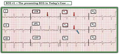
 

Figure-6: I've labeled Figure-1 to highlight ECG findings that suggest possible too-high placement of the V1,V2 electrode leads.


Final PEARL: Although one should clearly consider the possibility of previous “septal infarction” in the presence of deep Q waves (or QS complexes) in leads V1 and V2 — in the absence of another Q wave (or QS complex) in lead V3 — the vast majority of patients with Q waves in V1 and V2 but not V3, will not have prior “septal” infarction. 

  • Clinical correlation, comparison with prior tracings, and verification of correct lead placement would all be needed for optimally accurate comment on the likelihood of prior septal infarction in such cases.


==================================

Acknowledgment: My appreciation Sarah Johnson Saunders (from North Carolina, USA) for making me aware of this case and allowing me to use this tracing.

==================================


Related ECG Blog Posts to Today’s Case: 

  • ECG Blog #205 — Reviews my Systematic Approach to 12-lead ECG Interpretation (outlined in Figures-2 and -3, and the subject of Audio Pearl MP-23-LINK in Blog #205).
  •  
  • ECG Blog #269 — Reviews assessment of R-Wave Progression (including the causes of "Poor" R wave progression).
  •  
  • ECG Blog #203 — Reviews the basics for assessing Axis + a user-friendly approach for the ECG diagnosis of the Hemiblocks (with text and Video Pearls on these 2 subjects).
  •  
  • ECG Blog #234 — Reviews the ECG diagnosis of RVH and Pulmonary Disease.
  •  
  • ECG Blog #218 — Reviews when a T-Wave is Hyperacute.
  •  
  • The November 4, 2018 post in Dr. Smith's ECG Blog — from which the tracings in Figure-2 and Figure-3 of today's case are taken. 


Tuesday, January 4, 2022

ECG Blog #273 (73) — 30 Minutes After ED Arrival


A man in his 70s was seen in the ED (Emergency Department) for chest pain of several days duration. His initial ECG suggested an acute OMI (Occlusion-based Myocardial Infarction). Just 30 minutes later, while still in the ED — the patient became hemodynamically unstable, and the ECG in Figure-1 was obtained.


QUESTIONS:

  • How would YOU interpret the ECG in Figure-1?
  • How would you treat this patient?


Figure-1: ECG from a man in his 70s, who suddenly became unstable in association with this ECG. His initial ED ECG (done just 30 minutes earlier) — suggested an acute OMI.


=======================================

NOTE: Some readers may prefer at this point to listen to the 5:40-minute ECG Audio PEARL before reading My Thoughts regarding the ECG in Figure-1. Feel free at any time to refer to My Thoughts on these tracings (that appear below ECG MP-73).

=======================================


Today's ECG Media PEARL #73 (5:40 minutes Audio) — Reviews the concept of "Shark Fin" ST elevation and depression as a sign of extensive acute infarction.

  • NOTE: For more on "Shark Fin" ST elevation — Please see ECG Blog #265.


MY Approach to the Rhythm in Figure-1:

By the Ps, Qs & 3R Approach (which I reviewed in ECG Blog #185) — the rhythm in Figure-1 is fast and Regular — the ventricular Rate is between 190-200/minute — the QRS complex is wide (at least in some leads) — and, normal sinus P waves appear to be absent.

  • I can not rule out the possibility of atrial activity in the form of 1:1 VA (ie, retrograde) conduction — as a negative notch appears to be distorting the terminal part of the QRS complex (or the beginning of the ST segment) in each of the inferior leads. 
  • PEARL #1: Even if 1:1 retrograde P wave activity was present — this finding does not distinguish between VT (Ventricular Tachycardia) vs a reentry SVT (SupraVentricular Tachycardia), because both of these rhythms may conduct retrograde with a 1:1 VA ratio.


Is the QRS Complex WIDE in Figure-1?

My initial impression on seeing the ECG in Figure-1 — was that the QRS complex was definitely wide (The QRS certainly looks wide in leads V1, V2, and V3 — as well as in the remaining chest leads).

  • IF the QRS is indeed wide — then VT must be assumed until proven otherwise. This is because the rhythm would then be a regular WCT ( = Wide-Complex Tachycardia) at ~200/minute, without clear sign of sinus P waves — and statistical odds in favor of VT in this case would be over 90%! (See ECG Blog #220 — for full discussion of this subject).
  • That said — the QRS complex does not look wide in the 6 limb leads in Figure-1. This led me to question how certain I was that the rhythm was VT?


PEARL #2: Practically speaking — It does not matter IF the rhythm in Figure-1 is VT or some type of SVT rhythm. This is because we were told that the patient in today's case was hemodynamically unstable in association with this WCT rhythm at ~200/minute!

  • Since the patient is hemodynamically unstable because of the very rapid rate of this rhythm — immediate synchronized cardioversion is the treatment of choice regardless of whether this is VT or an SVT.


Comparing the First 2 Tracings:

The initial ECG for the patient in today's case ( = ECG #1) is shown in the TOP panel of Figure-2. We discussed this tracing in detail in ECG Blog #272. Pertinent findings included:

  • Sinus rhythm at ~70/minute — with normal intervals and a normal axis close to zero degrees. No chamber enlargement.
  • Low voltage.
  • A QS in lead III — and a tiny, fragmented QRS in lead aVF.
  • Hyperacute ST-T waves diffusely in the chest leads — with subtle-but-real reciprocal ST-T wave changes in the inferior leads. 
  • Clinical Impression of ECG #1: As noted in ECG Blog #272 — Given the history of chest pain over several days, I interpreted this tracing as suggestive of acute and/or ongoing extensive anterior infarction. Several of the markedly abnormal hyperacute chest lead T waves suggested a deWinter-like picture (See ECG Blog #183 — for discussion of deWinter-like T waves).


KEY Points in Comparison:

The reason for difficulty in determining whether the QRS complex in ECG #2 of Figure-2 is wide or not — is that a number of leads appear to manifest marked ST segment elevation that mimics QRS widening! That said — reasons why I interpreted ECG #2 as Ventricular Tachycardia until proven otherwise include:

  • The rate of the rhythm in ECG #2 (ie, ~200/minute) in this patient with hemodynamic instability. This rate is significantly faster than is generally seen for sinus tachycardia — and suggests that hemodynamic instability is likely to be because of the fast rate — which is indication for immediate cardioversion regardless of rhythm etiology.
  • The wide and predominantly negative initial part of the QRS complex in leads V4, V5 and V6 of ECG #2 — is very uncharacteristic for a supraventricular rhythm.
  • The almost completely negative QRS complex in each of the inferior leads in ECG #2. This suggests a very different (extreme) frontal plane axis during the tachycardia, compared to the relatively normal (at zero degrees) axis seen in ECG #1 during sinus rhythm.
  • Statistical odds strongly favor VT as the rhythm in ECG #2. The onus of "proof" lies with demonstrating  aberrant conduction — rather than the other way around. Since QRS morphology does not resemble any known form of bundle branch block — VT should be assumed until proven otherwise!


Figure-2: Comparison of the first 2 ECGs obtained in this case. ECG #1 is the initial ECG obtained in the ED (which was discussed in detail in ECG Blog #272). Just 30 minutes after arrival in the ED — ECG #2 was obtained. Is ECG #2 VT?


The Case Continues:

Shortly after ECG #2 was obtained — the rhythm deteriorated to VFib (Ventricular Fibrillation). The patient underwent an extended course of resuscitation — including intubation, multiple defibrillations and multiple medications (including epinephrine, lidocaine, amiodarone).

  • ROSC (Return Of Spontaneous Circulation) was finally achieved — and on stabilization, the patient was sent for cardiac catheterization. This revealed total occlusion of the mid-LAD  (Left Anterior Descending) coronary artery — which was stented. Minor lesions were noted elsewhere.
  • The post-resuscitation (after ROSC) ECG is shown in Figure-3. How would you interpret ECG #3 in light of the tachycardia seen in ECG #2? 


Figure-3: Comparison of the post-resuscitation tracing (ECG #3) — with the 12-lead tracing during the tachycardia (ECG #2)?


Interpreting the 2 ECGs in Figure-3:

It is easiest to first interpret the post-resuscitation tracing ( = ECG #3).

  • Sinus rhythm has been reestablished in ECG #3 at a rate of ~80/minute. The PR interval appears to be prolonged to ~0.24 second.
  • Once again — determination of QRS width is difficult. This is because of the tiny QRS amplitude in the limb leads, in which the QRS looks narrow. It is once again difficult to determine if the notching after the small R waves in inferior leads represents part of the QRS. Since the rhythm is sinus (upright P waves with a fixed PR interval in lead II) — this notch is unlikely to represent retrograde atrial activity.
  • The QRS does look wide in the chest leads — with terminal positivity in the anterior leads consistent with RBBB (Right Bundle Branch Block). In addition — there appears to be significant ST elevation in virtually all chest leads, which is most marked in leads V5 and V6. 
  • ST elevation is also seen in high-lateral leads I and aVL. Given how tiny QRS amplitude is in these 2 leads — the relative amount of ST elevation is marked.
  • There is suggestion of reciprocal ST depression in the inferior leads, probably with terminal T wave positivity.
  • Clinical Impression of ECG #3: This post-resuscitation ECG shows restoration of sinus rhythm — and confirms acute extensive anterior infarction with RBBB.


MY Thoughts:

Now that we know there is extensive ST elevation in the chest leads of the post-resuscitation tracing ( = ECG #3) — it becomes easier to appreciate that regardless of what the rhythm etiology of ECG #2 turned out to be — there was marked chest lead ST elevation in this tachycardia tracing that "blended in" with the terminal part of the QRS complex, making the QRS seem wider than it was. 

  • There was also indisputable ST elevation in leads I and aVL during the tachycardia (ie, in ECG #2).
  • Acknowledgment: Because of the difficulty determining the limits of the QRS complex in ECG #2 — I was not completely comfortable with my rhythm diagnosis of this tracing. I still favored VT for the reasons I cited earlier (ie, widened predominantly negative initial part of the QRS in V4-6 — the almost completely negative QRS in each of the inferior leads that is very different from the clearly upright QRS in the inferior leads of ECG #3 — and — statistics, that strongly favored VT, especially given the patient's compromised hemodynamic status).


PEARL #3: Sometimes, even with ventricular rhythms — you still will be able to detect acute ST elevation.

  • This is relevant clinically — since recognition that regardless of rhythm etiology, the very fast tachycardia in ECG #2 needs immediate treatment (ie, immediate cardioversion!).


Did YOU Notice the Shark Fin Morphology?

The reason today's case is so challenging — is the presence of "Shark Fin" morphology in both ECG #2 and ECG #3 (Figure-4).


Figure-4: I've added colored vertical lines to the 2 ECGs from Figure-3 — in order to highlight the end point of the QRS complex in each of the 12 leads (See text).


It's important to be aware of the pattern of "Shark Fin" morphology — in which the QRS complex looks wide, because it blends in with ST segments that show extreme ST elevation in multiple leads. As a result — the boundary between the end of the QRS complex and the ST segment becomes indistinguishable in those leads showing marked ST elevation or depression.

  • As discussed in detail in today's Audio Pearl (above) — "Shark Fin" ST segment elevation is most often a sign of severe transmural ischemia that results from acute coronary occlusion. Consideration of prompt cardiac cath is essential for clarifying the anatomy — since in many (most) cases, prognosis is likely to be poor unless there is prompt reperfusion.


PEARL #4: The KEY for confirming that "Shark Fin" morphology is the cause of the striking ECG picture this produces — is to find 1 or 2 leads in which you can clearly define the limits (end point) of the QRS complex. From there, with use of a simultaneously-recorded long lead rhythm strip — you can extrapolate where the end of the QRS will be in leads in which this boundary between the end of the QRS and the beginning of the ST segment is less clear. This concept is best illustrated in today's case by looking first at ECG #3 in Figure-4:

  • The most helpful leads for defining the end of the QRS in ECG #3 — are leads V4, V5 and V6. Note that I've drawn in a RED line parallel to the heavy ECG grid line in simultaneously-recorded leads V4,V5,V6. Note that I've extended this line down to the corresponding beat in the long lead II rhythm strip of ECG #3 (Figure-4).
  • The reason for continuing the RED line all the way down to the corresponding beat in the long lead II rhythm strip — is that this tells you where the QRS complex ends (and the ST segment begins) in the long lead II rhythm strip.
  • Knowing this landmark for the beats in the long lead II rhythm strip — allows us to draw in and extend upward the PINK lines parallel to the heavy ECG grid line in the other 3 sets of simultaneously-recorded leads (vertical PINK lines for ECG #3 in Figure-4).
  • Therefore: The post-resuscitation tracing ( = ECG #3) in Figure-4 shows restoration of sinus rhythm, with a widened qR pattern in lead V1 consistent with RBBB + massive (ie, "Shark Fin" morphology) ST elevation in leads V4,V5,V6. A lesser degree of ST elevation is seen in leads I, aVL; and in leads V1,V2,V3. Suggestion of subtle reciprocal ST depression is seen in the inferior leads. This strongly suggests acute proximal- or mid-LAD occlusion, with need for prompt cath.


What About "Shark Fin" Morphology in ECG #2?

Admittedly — I found it decidedly more difficult to delineate the boundary between the end of the QRS and the beginning of the ST segment in the tachycardia tracing ( = ECG #2 in Figure-4). This was because of the very fast rate and the very unusual appearance of  the QRS in the chest leads.

  • PEARL #5: Sometimes use of subsequent tracings allows retrospective interpretation of ECG findings that were not clear previously. So it is for assessment of ECG #2 in Figure-4. We have just established that there was massive (ie, "Shark Fin") ST elevation in leads V4,V5,V6 of ECG #3. Knowing this facilitates drawing in the vertical RED line in leads V4,V5,V6 of ECG #2.
  • Doing so defines the end of the QRS complex for the corresponding beat in the long lead II rhythm strip of ECG #2. Knowing this landmark for all beats in the long lead II rhythm strip of ECG #2 — allows us to draw in and extend upward the PINK lines parallel to the heavy ECG grid line in the other 3 sets of simultaneously-recorded leads (vertical PINK lines for ECG #2 in Figure-4).
  • Therefore: The tachycardia tracing ( = ECG #2) in Figure-4 shows a regular WCT rhythm with unusual QRS morphology, at a rate of ~200/minute — with what appears to "Shark Fin" ST elevation in at least several chest leads. Regardless of rhythm etiology — there is clearly significant ST elevation in leads I and aVL of ECG #2. Prompt cardioversion is essential!


How Well did ECG #1 Predict STEMI Location?

Comparison of the 2 tracings in Figure-5 provides another example of the concept I highlighted in Pearl #5. There is LOTS to be learned by going back after the conclusion of a case to reexamine data points along the way!

  • My clinical impression of ECG #1 had been that this tracing was suggestive of acute and/or ongoing extensive anterior infarction. I thought several of the markedly abnormal hyperacute chest lead T waves suggested a deWinter-like picture — that is typically associated with proximal- or mid-LAD occlusion in progress.
  • Given the extent of the marked ST-T wave abnormalities in ECG #1 — the possibility of deterioration with cardiac arrest should not be completely surprising. As we noted earlier — cardiac cath revealed acute occlusion of the mid-LAD, with minimal disease elsewhere. Comparison of ECG #1 with the post-resuscitation tracing ( = ECG #3) — shows dramatic evolution of the extensive anterior STEMI, with development of RBBB and marked ST elevation in those leads that previously manifested hyperacute T waves.
  • BOTTOM LINE: The ECG picture of hyperacute T waves in multiple leads of ECG #1 is an important one to remember! Unfortunately, the patient in today's case did not survive.


Figure-5: Comparison of the initial ECG in the ED — with the post-resuscitation ECG after ROSC (See text).


==================================

Acknowledgment: My appreciation to 유영준 (from Seoul, Korea) for making me aware of this case and allowing me to use this tracing.

==================================


Related ECG Blog Posts to Today’s Case: 

  • ECG Blog #185 – Reviews the Ps, Qs, 3R Approach to Systematic Rhythm Interpretation.
  •  
  • ECG Blog #220 — Reviews the approach to a patient with a regular WCT ( = Wide-Complex Tachycardia) rhythm.

  • ECG Blog #272 — Reviews the initial ECG in Today's Case.
  •  
  • ECG Blog #205 — Reviews my Systematic Approach to 12-lead ECG Interpretation (outlined in Figures-2 and -3, and the subject of Audio Pearl MP-23-LINK in Blog #205).

  • ECG Blog #265 — Reviews the concept of "Shark Fin" ST elevation as a sign of extensive acute infarction.
  •  
  • ECG Blog #193 — illustrates use of the Mirror Test to facilitate recognition of acute Posterior MI. This blog post reviews the basics for predicting the "culprit artery". NOTE: The Audio Pearl reviews the concept of why the term "OMI" ( = Occlusion-based MI) should replace the more familiar term STEMI.
  •  
  • ECG Blog #218 — Reviews of the concept of  WHAT is a "Hyperacute" T Wave (Also see the Audio Pearl in this post).
  •  
  • ECG Blog #183 — Reviews a case of deWinter-like T Waves (with the Audio Pearl in this post discussing some variants of the deWinter T wave pattern). 
  • ECG Blog #53 — Reviews another case of deWinter T Waves.

  • ECG Blog #258 — Reviews the concept of HOW to "Date" an Infarction (Also see the Audio Pearl in this post).