Sunday, December 23, 2018

ECG Blog #158 — Acute "Indigestion" ...

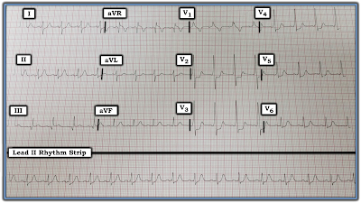
Imagine the ECG in Figure-1 was obtained from a previously healthy, middle-aged man who presented to an ambulatory clinic for “indigestion”.
  • How would you interpret this ECG in light of the above history?
  • Are there acute changes?

Figure-1: How to interpret this ECG if the history is "acute indigestion"?

========================

Interpretation: The rhythm for the ECG shown in Figure-1 is sinus at a rate of ~70/minute. All intervals are normal. The mean QRS axis is normal (about +30 degrees — as judged by the upright QRS in leads I and aVF, with greater net positivity in lead in lead I). There is no chamber enlargement. Regarding assessment of Q-R-S-T Changes:
  • A large Q wave is seen in lead III — and small q waves are present in leads II and aVF.
  • Transition is normal (occurs between V3-to-V4).
  • There is coved ST elevation in leads III and aVF — with a hint of early T wave inversion in these leads. There also appears to be slight-but-real ST elevation in lead II (See below).
  • ST depression is seen in lead I (slightly), and in lead aVL (more definite) — and probably also in lead V2.
  • The T wave is relatively flat in leads V5 and V6 (normally the T wave is clearly upright in these lateral chest leads).

IMPRESSION: The combination of findings described above suggests we should assume inferior MI (Myocardial Infarction) has occurred at some point, most probably in the recent past — at least, until proven otherwise. The challenge is to determine the probable age of this MI. We highlight the following points:
  • PEARL #1: I like using the concept of “patterns of leads” — especially when assessing the ECG of a patient suspected of acute ischemic heart disease. To apply this concept — Look at all leads that view a given area of the heart at the same time. Thus, we look here at the 3 inferior leads (II,III,aVF) at the same time (Figure-2). Given that 2 of these leads (ie, leads III and aVF) clearly show Q waves + ST elevation + beginning T wave inversion — it becomes exceedingly likely that the much more subtle findings in lead II are also real. These findings in lead II consist of ST elevation (RED arrow pointing to the ST segment which lies above the RED horizontal baseline in this lead) — and — and the small q wave (within the RED circle).

  • PEARL #2: With acute coronary disease — an almost "magical" reciprocal ( = mirror-image) relationship will usually be seen between ST-T wave deviations in leads III and aVL. To illustrate this concept — I have placed the mirror-image of the first 2 beats in lead III just over the complexes in lead aVL — and, the mirror-image of the first 2 beats in lead aVL just over the complexes in lead III. Recognition of this “almost magical” reciprocal relationship (as we see here in Figure-2) — virtually confirms acute (or at least recent) changes of MI.
  • Reciprocal ST flattening/depression is also seen in leads I and V2 in Figure-2 — albeit quite modest in degree. In the setting of acute or recent inferior MI — ST depression in anterior leads (ie, V1, V2 and/or V3) typically indicates posterior wall involvement — so we suspect infero-postero MI in this case.

  • PEARL #3: It is uncommon for the amplitude of a positive T wave in lead V1 to be greater than the amplitude of the positive T wave in lead V6. When this relationship is seen in a patient with potentially acute symptoms (as is the case in Figure-2) — acute ischemia is likely.

  • NOTE — There may also be RV (right ventricular) involvement in this case. Lead V1 is both an anterior, as well right-sided lead. Proximal RCA (Right Coronary Artery) occlusion often results in acute infarction of the 3 areas of the heart supplied by the RCA, which are the inferior and posterior wall of the LV (left ventricle), and the RV (right ventricle). Diagnosis of acute RV MI is often challenging on a standard 12-lead ECG — because the expected right-sided ST elevation is frequently cancelled out by anterior ST depression from associated posterior MI. While we might suspect this as the reason for the modest amount of anterior ST depression and the upright T wave in lead V1 of Figure-2 — the only way to know if there is RV involvement in this case would be with right-sided leads (ie, leads V1R-thru at least V4R).

Figure-2: We have labeled Figure-1 to highlight subtle changes in lead II — and the “magic” reciprocal (mirror-image) relationship with acute or recent coronary disease between leads III and aVL (See text).

========================

BOTTOM Line: The ECG in Figure-2 strongly suggests that there has been an Infero-Postero MI (possibly also with RV involvement). But the question remains as to whether this is acute (ie, within the last couple of hours) — recent (within the past day or 2 or 3) — or more remote than just a few days ago?
  • Treatment recommendations for this patient will vary dramatically depending on the acuity of the MI. For example, immediate cardiac catheterization and/or thrombolytic therapy are less likely to be helpful if much time has passed since the acute event.
  • The History in this case is very important. Because the area of the heart viewed by the inferior leads (especially leads III and aVF) lies on top of the diaphragm — it is not at all uncommon for patients with inferior MI to present with GI symptoms. If this is in fact the case here — it will be important to try to elicit from the history WHEN this patient’s “indigestion” began.
  • ECG findings in Figure-1 that suggest a more acute STEMI (= ST Elevation MI ) are: i) that there is ST elevation in each of the inferior leads; ii) that there is reciprocal ST depression, including that “magical” mirror-image picture for the ST-T waves in leads III and aVL; and, iii) that T wave inversion looks like it might only be beginning ...
  • On the other hand, ECG findings that suggest a less acute picture include: i) that the overall amount of ST elevation is relatively modest; ii) that a very large Q wave has already formed in lead III; iii) that the amount of reciprocal ST depression is also extremely modest; and, iv) that the patient presented to an ambulatory clinic instead of to an ED (Emergency Department). 
  • While it certainly occurs that patients with acute MI can present to an ambulatory clinic — I’ve observed over the years a “self-selection” process, whereby statistical likelihood of an acute event seems greatly enhanced when patients call 911 and present with their symptoms to the ED instead of to an outpatient clinic.

=============================

My BEST Guess: Pending additional historical information — the seemingly uncertain onset of GI complaints + outpatient presentation + an ECG with a very large Q wave in lead III + relatively modest amount of ST elevation and depression all suggest a less acute onset for this MI. Whether this dates the infarct as having occurred 12-24 hours ago, or even longer ago than that — is uncertain from the information available.
  • That above said, one can not rule out a more acute onset — especially if the reason for relatively little ST elevation is spontaneous reperfusion (in which case, the risk of potential reocclusion may be considerable).
  • To emphasize — clinical priorities and the treatment approach would be very different IF the history suggested a recent and/or ongoing onset. For example, prompt cath to define the anatomy would be indicated if the patient had called 911 and/or presented to the ED with new-onset chest pain ... Sometimes, "Ya gotta be there" to fully appreciate the clinical situation.
  • In Summary — prompt assessment and more information is needed to determine true risk of this patient. This most likely would (should) include timely cardiac catheterization to define the anatomy.

-------------------------------------------------------------------------
-------------------------------------------------------------------------
  • NOTE: For more on our approach to dating an infarct — Please see our ECG Blog #142.


Saturday, December 15, 2018

ECG Blog #157 (RBBB - WPW - RVH - Post MI)

The ECG in Figure-1 was obtained from a 35-year old man with a 20-pack year history of smoking — who presented with new-onset chest discomfort. He was hemodynamically stable at the time this tracing was obtained. 
  • How would you interpret his ECG?
  • Is there RBBB?
  • Should the cath lab be activated?

Figure-1: ECG obtained from a 35-year old man with new-onset chest discomfort. Should the cath lab be activated?


=============================
Interpretation: There is a regular sinus rhythm in Figure-1 at a rate of ~90/minute. At first glance, the PR interval looks normal — but the QRS complex looks wide, with a pattern in lead V1 that suggests RBBB (Right Bundle Branch Block). That said, this is not RBBB. Why not?
=============================


ANSWER: The pattern of QRS widening with the upright complex in lead V1 is not the result of RBBB. The reason for this is highlighted by the RED arrows in Figure-2, which point to delta waves that are evident in leads I and V2-thru-V5. This patient has WPW (Wolff-Parkinson-White) Syndrome.
  • Delta waves are not obvious in a number of leads on this tracing. This is especially true in several of the limb leads. It was only after I saw unmistakable slurring of the initial part of the QRS complex in certain leads (most evident in leads V2, V3 and V4) — that I realized this patient has WPW.
  • PEARL #1: Delta waves are not always seen in all 12 leads of the ECG when a patient has WPW. As a result, it can sometimes be quite challenging to make the diagnosis. This problem is complicated by the reality that some patients (more common in older individuals) normally manifest some initial slowing (and therefore slurring) of the early part of the QRS complex. BOTTOM LINE: When contemplating the possibility of WPW — all 12 leads should be carefully scrutinized. If questionable initial slurring is minimal and only noted in 1 or 2 leads — then the patient probably does not have WPW. But if unmistakable delta waves associated with a short PR interval are definitely seen (as is highlighted by the RED arrows in leads V2, V3 and V4 of Figure-2) — then you have made your diagnosis despite the absence of obvious delta waves in some of the other leads.
  • PEARL #2: Once you determine the patient has WPW — you’ll often be able to GO BACK and recognize subtle deflections that in retrospect are also delta waves. For example, in Figure-2 — I initially thought there was a Q wave in lead III (WHITE arrow). However, the fact that this negative deflection in lead III occurs at precisely the same instant in time as the delta wave in simultaneously-obtained lead I (dotted WHITE line, leading up to the RED arrow in lead I) — tells us that this negative deflection in lead III is actually a negative delta wave.
  • This illustrates why WPW is often referred to as, “the great ECG mimic” — because WPW may simulate a number of ECG findings, including myocardial infarction (if there are multiple negative delta waves), as well as chamber enlargement, ischemia, and conduction defects! Thus, there is no RBBB in Figure-2 — because this patient has WPW.
  • Other examples of “retrospective recognition” of delta waves in Figure-2 are seen in leads II, aVL, and aVF. Thus, the suggestion of a tiny “extra little bump” on the baseline in leads II and aVF probably represents subtle delta waves that are almost entirely isoelectric.

Figure-2: We have labeled Figure-1 to explain why there is no RBBB. But what about the ST depression within the WHITE rectangles? Should the cath lab be activated? NOTE: Although the ECG is not straight, and the grid lines are slanted — we have drawn the dotted WHITE line parallel to the heavy grid line just before it, to show simultaneously-occurring events in leads I, II and III. (See text).


=============================
ECG Findings with WPW: When the rhythm is sinus — conduction of the electrical impulse in a patient with WPW begins as usual in the SA node. From there, the impulse may be conducted to the ventricles in one of 3 ways:
  • Possibility #1: Entirely over the AP (Accessory Pathway) — in which case the AV node will be completely “bypassed”, resulting in: i) a delta wave (due to slow conduction through nonspecialized myocardial tissue); ii) a short PR interval (since the usual delay in conduction inherent within the AV node is bypassed); and, iii) a wide QRS complex (from slowed conduction through the ventricles) — OR —
  • Possibility #2: Conduction to the ventricles with WPW may at times be entirely over the normal AV nodal pathway (in which case there will be no delta waves on the ECG; the PR interval will be normal; and the QRS complex will be narrow and look normal) — OR —
  • Possibility #3: There may be simultaneous conduction over both the AP and the normal AV nodal pathway! If relatively more conduction occurs over the AP — then the QRS complex will be wide, and there will be delta waves and a short PR interval. If instead, preexcitation is limited so that conduction occurs primarily over the normal AV nodal pathway — then the features of WPW may be subtle, and you may not even realize that an AP exists. But, if a significant amount of conduction is simultaneously transmitted over both the AP and the normal AV nodal pathway — then the resultant PR interval and QRS complex may reflect features intermediate between normal and preexcited complexes.
=============================
NOTE: For more on the ECG features of WPW, as well as clinical implications when WPW is found in an asymptomatic patient — Please see our ECG Blog #153.
=============================

QUESTION: What about the marked ST segment depression seen within the WHITE rectangles in Figure-2 (BLUE arrows)? Is this ST depression that is seen in most chest leads reflective of ischemia in this 35-year old man with new chest discomfort? — OR — Are we unable to assess the significance of this finding because this patient has WPW?
  • KEY POINT: In general — assessment of ST-T wave deviations (elevation or depression) do not reliably predict underlying ischemia or infarction when there is considerable preexcitation. This is because since the sequence of depolarization is altered by WPW — the sequence of repolarization (and therefore the appearance of ST-T waves) will also be altered. That said, the shape and amount of ST depression within the WHITE rectangles in Figure-2 is so pronounced — that one wonders if despite the presence of WPW, this ST depression might nevertheless indicate acute ischemia?

=============================
Follow-Up: The cardiologist involved in the case was as uncertain as I am looking at this ST depression (within the WHITE rectangles) — as to whether it is a marker of acute ischemia, or simply an accompaniment of this patient’s WPW. Given this patient’s history ( = risk factors + new-onset chest pain) — cardiac catheterization was performed. The coronary arteries were free of significant disease.

-------------------------------------------------------------------------
COMMENT: Most of the time, you will not be able to appreciate ischemia or acute infarction on the ECG of a patient with WPW. That said, on occasion — ST-T wave changes may be so marked as to suggest acute disease, necessitating cardiac cath to define the anatomy. This was the situation in this case.

-------------------------------------------------------------------------
Acknowledgment: My thanks to Dr. Ahmed Abbas from Amman, Jordan, for his permission allowing me to use this tracing and clinical case.

-------------------------------------------------------------------------
-------------------------------------------------------------------------
  • NOTE: For more on the ECG features of WPW, as well as clinical implications when WPW is found in an asymptomatic patient — Please see our ECG Blog #153.
  • CLICK HERE — for a link to Dr. Stephen Smith's ECG Blog post from March 1, 2012, in which he shows a series of WPW tracings which either mimic, hide or reveal acute MI.


Sunday, November 18, 2018

ECG Blog #156 (LVH – RVH – LAA – RAA – Strain – Ischemia)

The ECG shown in Figure-1 was obtained from a 40-year old man. Without the benefit of any history — How would you interpret this tracing?
  • Is there evidence of an acute coronary syndrome?
  • Is there a common diagnosis that potentially explains all of the findings?

Figure-1: ECG obtained from a 40-year old man. How would you interpret this tracing? (See text). 


=========================================
Interpretation: There is baseline artifact that is most marked in lead V1. That said — this tracing is definitely interpretable. Approaching the ECG in Figure-1 systematically — We note the following:
  • Rhythm & Rhythm — The rhythm is sinus at a rate just under 100/minute.
  • Intervals — The PR, QRS and QT intervals are all normal (ie, the PR interval is not more than 1 large box — QRS duration is not more than half a large box — and despite the fairly rapid rate, the QT interval is not more than half the R-R interval). NOTE: We review our rapid assessment approach for intervals in ECG Blog #89.
Axis — There is marked RAD (Right Axis Deviation) — as determined by predominant negativity in lead I, that occurs in association with a predominantly positive QRS complex in lead aVF.
  • NOTE: Although the finding of a predominantly negative QRS complex in lead I (especially in association with a completely negative QRS in lead aVL) should prompt consideration of lead misplacement — this is not the explanation here. Instead, the fact that the P wave and T wave in lead I are both positive, in conjunction with the expected negative P wave in lead aVR — tells us there is no lead misplacement. Instead, there is simply marked RAD.
Chamber Enlargement — As assessment for chamber enlargement highlights the most important findings in this case — we dissect our approach:
  • We review assessment for atrial abnormality in ECG Blog #75. Definite criteria for RAA (Right Atrial Abnormality) are met in Figure-1 — in that the P wave is very tall and peaked in each of the 3 inferior leads (the P wave is at least 3mm tall in lead II). In addition, P waves are even taller, as well as peaked and pointed in 5 out of the 6 chest leads (ie, leads V2-thru-V6). We emphasize that in the vast majority of cases, ECG diagnosis of RAA is made from assessment of the limb leads. The ECG in Figure-1 provides one of those rare instances in which P wave appearance in the chest leads assists to confirm our impression of RAA.
  • PEARL: The presence of a very pointed P wave in either lead V1 and/or V2 is an insensitive, but highly specific ECG finding in favor of RAA that most often indicates marked RA enlargement and/or significant pulmonary hypertension. This is especially true when P wave amplitude of these pointed chest lead P waves is excessive, as it is here (It is rare indeed to see 5mm P waves in any ECG lead!).
  • Although one might think that the deep negative component to the P wave in lead V1 indicates LAA (Left Atrial Abnormality) — we suspect instead, that this is simply further evidence in support of marked RAA. That’s because on occasion, the RA (right atrium) may enlarge so extensively, that it “flops over” and is viewed by right-sided lead V1 as producing a wave of depolarization moving away from V1. The fact that the negative component to the P wave in lead V1 is so narrow (nearly the mirror-image of the upright pointed and very skinny P wave in lead V2) — makes us suspect that this is what is happening here.
  • We review assessment for RVH (Right Ventricular Hypertrophy) in ECG Blog #77. As emphasized in Blog #77 — we view the ECG diagnosis of RVH as a "Detective" Diagnosis — since rarely will any one single finding clinch the diagnosis. Instead — determination of RVH is usually made by deduction from identifying a combination of suggestive ECG findings. Among the findings we note in Figure-1 that suggest RVH include: i) Marked RAD; ii) Marked RAA (since it is rare for RVH not to be present when there is right atrial enlargement); iii) Predominant R wave in lead V1 (Normally the QRS should be predominantly negative in lead V1); iv) Persistence of S waves through to lead V6 (Normally the QRS complex is all positive by the time one reaches lead V6); and, v) Diffuse T wave inversion suggestive of RV "Strain" (See below!).
  • PEARL: There is actually a small q wave in lead V1, prior to the predominant R wave in this lead. In patients with RVH — this initial small q wave in lead V1 is often associated with pulmonary hypertension.
Q-R-S-T Changes — The last part of our systematic interpretation entails assessment of Q-R-S-T wave changes.
  • Q Waves — We have already noted the QS complex in lead aVL, and the small q (qR complex) in lead V1. There is also a Q wave in lead aVR which is common, and not indicative of anything special.
  • R Wave Progression — We have also already noted predominant positivity of the QRS complex in lead V1.
This leaves us with assessment of ST-T Wave Changes — which show ST-T wave depression in multiple leads!
  • PEARL: There are 2 lead areas that typically manifest asymmetric ST-T wave depression with RV “Strain”. These are the inferior leads (ie, II,III,aVF) — and the anterior leads (usually V1,V2,V3). RV “strain” may be seen in one or both of these areas. Sometimes when there is marked RV “strain” — ST-T wave depression may extend further across precordial leads. This appears to be the situation in Figure-1.
CLINICAL IMPRESSION — While one might be tempted to ascribe the diffuse ST-T wave depression in Figure-1 to ischemia — it is far more likely to reflect marked RVH with Pulmonary Hypertension. That’s because this unifying diagnosis potentially explains all of the important ECG findings we have noted above = marked RAD — RAA — the qR pattern and predominant R wave in lead V1 — persistence of S waves through to lead V6 — and diffuse ST-T wave depression consistent with RV “strain”.
  • Clinical Correlation — This 40-year old man has uncorrected Tetralogy of Fallot. This explains the findings of right atrial and right ventricular enlargement, with pulmonary shunting and pulmonary hypertension.
  • For brief review of the principal clinical findings associated with Tetralogy of Fallot — CLICK HERE.
=========================================
Acknowledgment: My thanks to Haval Issa from Duhok, Iraq for his permission allowing me to use this tracing and clinical case.
=========================================


Sunday, September 23, 2018

ECG Blog #155 (PACs – Wandering – MAT )


The ECG in Figure-1 was obtained from a woman in her 50s, who complained of intermittent chest discomfort in recent weeks. She was hemodynamically stable at the time this tracing was obtained.
  • How would you interpret her ECG?
  • HINT: The rhythm is not sinus ... 

Figure-1: ECG obtained from a woman in her 50s, with intermittent chest discomfort. How would you interpret this tracing?


Interpretation: The interesting part of this tracing is the rhythm. We have numbered the long lead II rhythm strip. What do you see?


-------------------------------------------------------------------------
ANSWER: The rhythm is irregular. The QRS complex is narrow. P waves are present — albeit as you look from 1 beat to the next in the long lead II rhythm strip, it should be clear that P wave morphology changes ...
  • We identify 3 different shapes of P waves on this tracing (Figure-2). That is, P waves are tall and pointed in front of beats #1,5,6,7 and 8 (RED arrows) — P waves are round in front of beats #2, 9 and 10 (BLUE arrows) — and, P waves are pointed, but not quite as tall in front of beats #3,4,11,12 (GREEN arrows).
  • Each of the P waves in this long lead II is conducting. We know this because for each of these 3 different P wave shapes — all beats of that shape manifest the same PR interval.

Figure-2: Colored arrows highlight atrial activity in the long lead II rhythm strip. What is the rhythm? (See text).


The principal differential diagnosis for an irregular rhythm with different-shape P waves that are conducting is: i) Sinus rhythm with multiple PACs; ii) MAT (Multifocal Atrial Tachycardia); and, iii) Wandering pacemaker. Regarding this differential:
  • This is not sinus rhythm with PACs — because there is no predominant underlying sinus rhythm. And, although R-R intervals are shorter in some places — there are no beats that are especially early (ie, there are no PACs).
  • This is not MAT — because P wave morphology and the PR interval do not change from one-beat-to-the-next. Instead, there is gradual change in the site of the supraventricular pacemaker over the course of several beats. This strongly suggests the presence of a wandering atrial pacemaker.

-------------------------------------------------------------------------
What is a Wandering Pacemaker?
Occasionally — the site of the atrial pacemaker may shift (wander) away from its usual site of origin in the SA (Sino-Atrial) Node. In most cases — wandering atrial pacemaker is a benign normal variant that occurs in patients without underlying heart disease. It may result from variationsin vagal tone (that slow SA nodal discharge and allow other atrial sites to temporarily emerge) — or there may be no obvious cause.
  • ECG recognition of wandering pacer requires a long enough rhythm strip to appreciate gradual change over a period of beats from one P wave morphology to another. Technically, there should be at least 3 different atrial sites — in order to distinguish a wandering atrial pacemaker from a simple atrial escape rhythm. The clinical reality, is that most of the time — the period of monitoring available for our scrutiny will simply not be long enough to appreciate gradual shift in the site of the atrial pacemaker to at least 3 different sites. As a result, true wandering pacemaker is not a common diagnosis.
  • KEY: All too often, even experienced interpreters fail to adequately assess the long lead rhythm strip. On seeing a few upright P waves — the “eye” tends to assume that the rest of the rhythm is also sinus. All it takes to avoid missing subtle deviations from strict sinus rhythm is a few careful seconds in which you ensure that you scrutinize the P wave preceding each QRS complex in the long lead rhythm strip. You’ll be amazed at how disciplined addition of these few extra seconds will easily pick up those cases in which the rhythm is not strictly sinus.
  • P.S.: Other than the rhythm — there are minimal nonspecific ST-T wave abnormalities in several leads that do not appear to be acute. These include shallow T wave inversion in lead III, and relative ST-T wave flattening in leads II, aVF, V5, V6. Thus, the wandering pacemaker is likely to be unrelated to the cause of this patient's intermittent chest discomfort.
==================================
COMMENT: The example of wandering atrial pacemaker in Figure-2 is unique in our experience for allowing definitive diagnosis of this rhythm in no more than a short long lead II rhythm strip. Most of the time, a much longer period of monitoring is needed.

==================================
Acknowledgment: My thanks to Joshua Wyeth from Petoskey, MI, for his permission allowing me to use this tracing and clinical case.
==================================

Related ECG Blog Posts to Today's Case:
  • ECG Blog #65 — for more on distinction between MAT, Wandering Pacer & Sinus Rhythm with PACs.
  • ECG Blog #200 — for more on distinction between these rhythms (plus the 4-minute Audio Pearl in this blog post).